
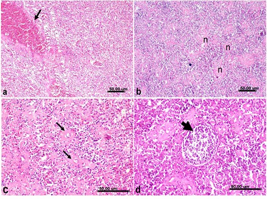
Figure 7
Histological sections of spleen from H5N8 infected duck, showing a) Severe congestion of splenic vessels (arrow) and sinusoids b) Spleen showing severe lymphocytic depletion with marked necrosis of lymphoid follicles (n). c) Fibrinoid necrosis of splenic arterioles(arrow). d) lymphocytic depletion and lymphocytolysis of lymphoid elements comprising the bursal associated follicles (arrow). (Stain, H&E).