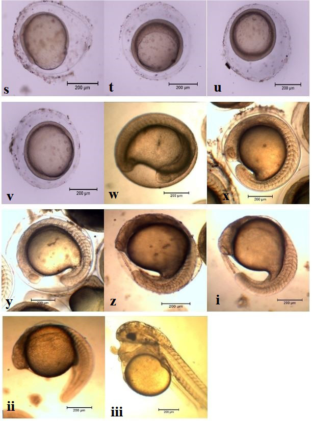
Figure 2:
Development stages of M. gulio (a) Unfertilized egg, (b) Fertilized egg, (c) Blastodisc, (d) 2-cell stage, (e) 4-cell stage, (f) 8-cell stage, (g) 16-cell stage, (h) 32- cell stage, (i) 64-cell stage, (j) 128-cell stage, (k) 256-cell stage, (l) 512-cell stage, (m) Oblong stage, (n) Sphere stage, (o) Morula stage, (p) Blastula stage, (q) 60% epiboly, (r) 70% epiboly, (s) 80% epiboly, (t) 90% epiboly, (u) Epiboly complete, (v) Bud stage, (w) Somites formation, (x) Segmentation-1, (y) Segmentation-2, (z) Segmentation-3 (i) Just before hatching, (ii) Newly hatched, (iii) larvae 24 hr larvae.