
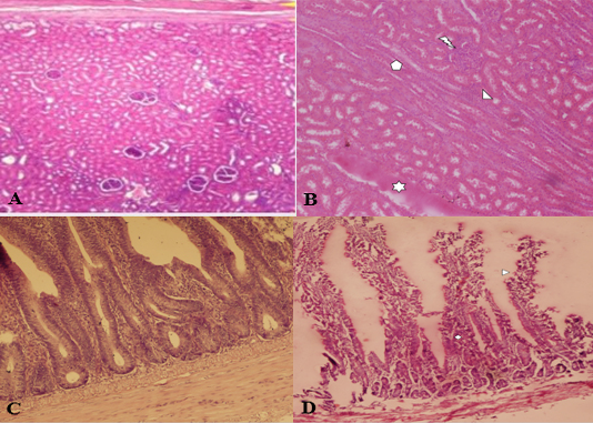
Fig. 1.
Histological structure of sheep kidney (A, B) and intestine (C, D). Control kidney (A), sheep kidney infected with C. perfringens type D (B) showing leukocyte infiltration, cast deposition, tubular epithelial cells and severe degeneration in tubular epithelium. Control intestine (C), intestine of infected sheep (D) showing sloughing of villi and leukocytic infiltration (D). **Stain: H&E (Hematoxcillin and Eosin); Magnification: A, B, 100x; C, D, 400X.