
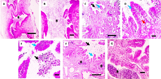
(A and B) Epididymis, Gp1 control showing epididymal duct with great quantity of spermatozoids (asterisk) in lumen and normal epithelial lining (white arrow), (C, D and E), Gp2 showing marked decrease in diameter of epididymal duct and amount of spermatozoids in lumen with presence of large amount of exfoliated germ cells and round bodies in the lumen (black arrows), hyperplastic alteration in the lining epithelium forming infolding or cribriform change and pseudoglandular structures (blue arrows) besides hyperplasia of clear cells (red arrow). (F and G), Gp3 showing slight increase in amount of spermatozoids in lumen (asterisk) with slightly decreased hyperplastic alteration in the lining epithelium (blue arrows) besides presence of few exfoliated germ cells and round bodies in the lumen (black arrows). H and E, Low magnification X: A, C and F: 100 bar 100 and high magnification X: B, D, E and G:400 bar 50.