
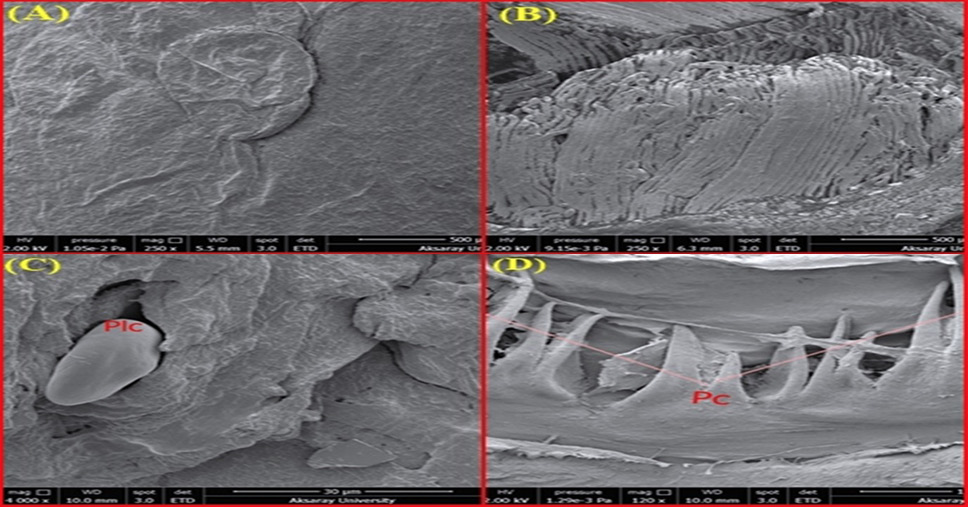
Fig. 6.
(A) SEM view of the surface epithelium of the corpus portion of the tongue (9-13 weeks). (B) Cross-sectional SEM view of longitudinal tongue muscles (6-7 weeks), (C) SEM view of the surface of the tongue body and papilla linguales caudales (6-7 weeks), (D) SEM view of the papillae of the radix part of the tongue (6-7 weeks), (Pc) Papilla conicae.