
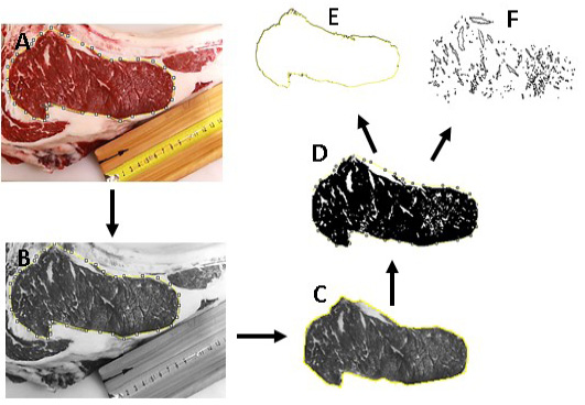
Figure 1
Image analysis method analysis using J software for estimating marbling percentage. The longissimus thoracis muscle cross-section (rib eye area) was firstly selected (A), converted into 8-bit image (B) and then the selected area was separated (C) and binarized (D) to highlight the marbling fat (white spot particles) over muscle background (black). Then, the binarized image was separated into two images; one showing the whole selected rib eye area (E) and one presenting spots areas of spotted particles (F). Marbling percentage was calculated as the marbling particles area (F) related to the area of the selected rib-eye area (E).