
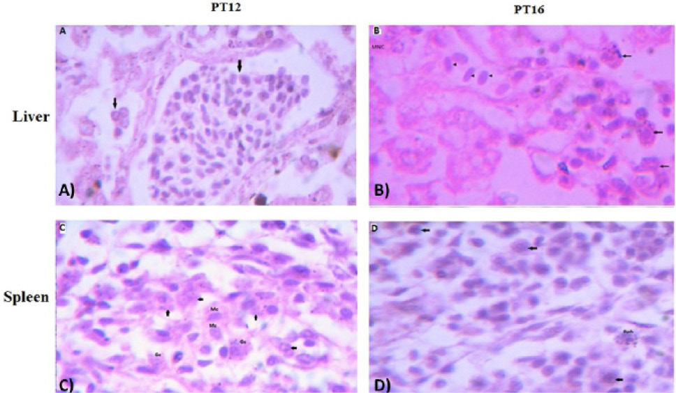
Fig. 2.
The histological examination of liver (upper panel) and spleen (lower panel) tissues of aHEV positive layer chickens PT12 and PT16; (A) PT12 liver histology showing lymphocytic infiltration in liver tissues causing portal phlebitis and periphlebitis (arrows) ; (B) PT16 liver histology with lymphocytic infiltration (arrow heads) ; granulocytes (arrows) and multifocal necrosis (MNC); (C) PT12 spleen histology showing infiltration of monocytes (Mc); granulocytes (arrows); (D) PT16 spleen histology showing infiltration of basophils (Bph) and other large cells (arrows). The images were observed under 100X magnifications.