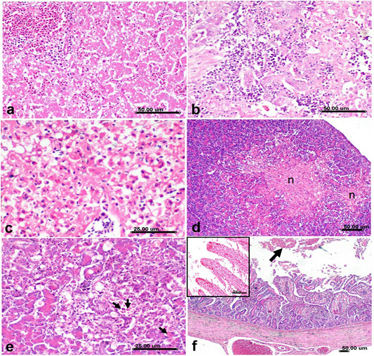
Histological sections of Liver, pancreas and duodenum from H5N8 infected duck, a) Liver showing severe congestion of portal vessels and hepatic sinusoids associated with vacuolization of hepatocellular cytoplasm. b) Liver showing lymphocytic infiltration of hepatic sinusoids. c) Liver showing severe necrotic reaction of hepatocytes with karyorrhexis and pyknosis of their nuclei d) Pancreas showing multifocal necrosis of exocrine pancreatic acini that replaced with eosinophilic necrotic tissue debris (n). e) Pancreas showing vacuolization of pancreatic acinar epithelium associated with apoptosis(arrow). f) Duodenum showing shortening, atrophy and blunting of duodenal villi, severe congestion of submucosal and serosal vessels and mucosal luminal hemorrhage (arrow), the inserted box in upper left corner showing severe ulceration of mucosa with congestion of blood capillaries in lamina propria that was infiltrated by mononuclear cells. (Stain, H&E).