
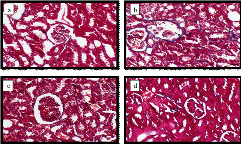
Fig. 7.
Light micrograph of kidney, (a) control animals showing thin layer of periglomerular and peritubular fibrous layer (arrow); (b) PAT -treated animals showing increased the thickness of the periglomerular and peritubular fibrous layer (arrow); (c) PAT and ginger -treated animals for 4 weeks showing thin periglomerular and peritubular fibrous layer (arrow) and (d) PAT and ginger -treated animal for 8 weeks showing thin perivascular layer of fibrous connective tissue (arrow), Masson’s trichrome stain, bar= 40 µm.