
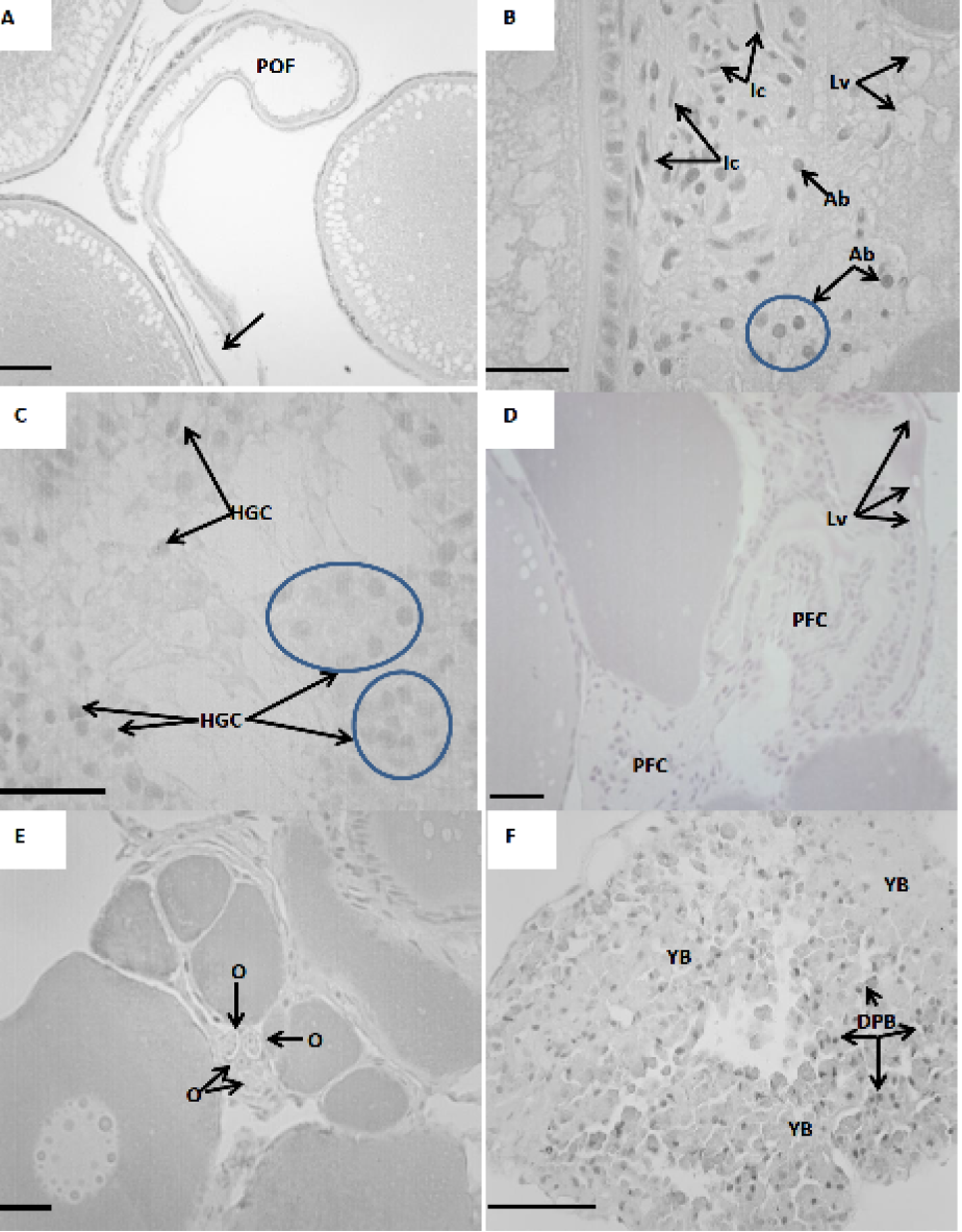
Fig. 4.
Atresia at post ovulatory follicle of C. Gariepinus. A, arrow shows evidence of the rupture of the follicular wall after ovulation; B, formation of interstitial cells; C, hypertrophy of granulosa cells are shown; D, phagocytıc follicular cells are shown; E, Arrow shows oogonia which were occasionaly detected amongst the connective tissue; F, arrows show yellow bodies which prominently represented in post ovulated ovaries. Dark pigmented bodies are also shown. POF, post ovulatory follicle; YB, Yellow bodies; Ab, apoptotic bodies; O, oogonia; DPB, dark pigmented bodies; HGC, hypertrophied granulosa cells; PFC, phagocytic follicular cells; Lv, large vacuole; Ic, interstitial cells. Scale bar, 200µm. Stain: hematoxylin & eosin.