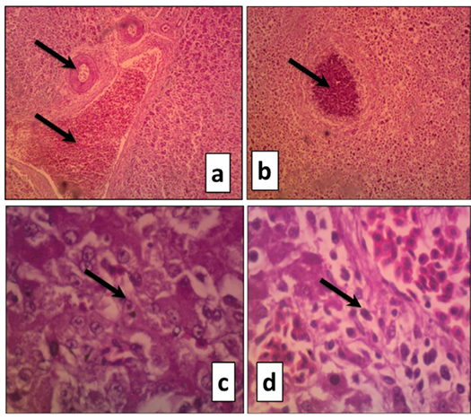
Figure 4
Liver sections of duckling naturally infected with DVH (H&E) showing
a. Severe portal vein congestion (arrow). The bile duct suffered from narrowing because of epithelial lining hypertrophy accompanied by severe hepatocyte necrosis (X100)
b. Focal area of coagulative necrosis characterized by inflammatory cells infiltration (arrow) accompanied byhepatic cord disorganization and severe hepatocytes necrosis (X200)
c. Large eosinophilic intranuclear inclusion surrounded by a clear halo in the hepatocytes (X400)
d. Large eosinophilic intranuclear inclusion surrounded by a clear halo in the hepatocytes (X400)