
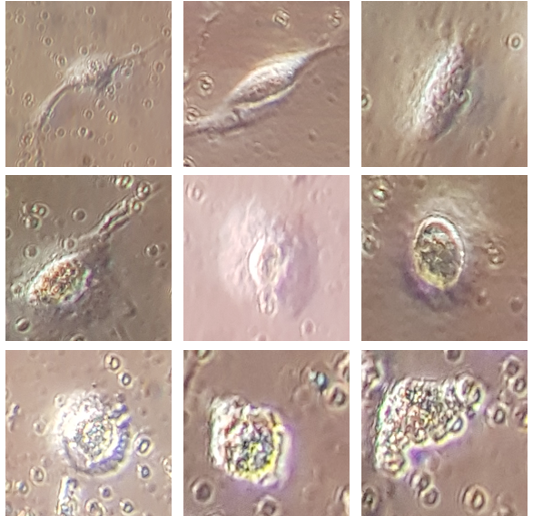
Figure 4
Micro-photos for vNDV effect on MSCs-CD105 during inhibition process, A) single cell shown, a complete external membrane without any damage without virus treatment, B) the beginning of a decrease in the terminal arms of cells immediately after treatment with the virus, C) the beginning of cells are vesicles, D) nods appearing on the outside of the cells, E) swelling and increase in cell size and giant cell formation, F) increasing in swelling, G) beginning in explosion, H) unstable cell, I) cell lysis and degradation, magnification power equal 100x.