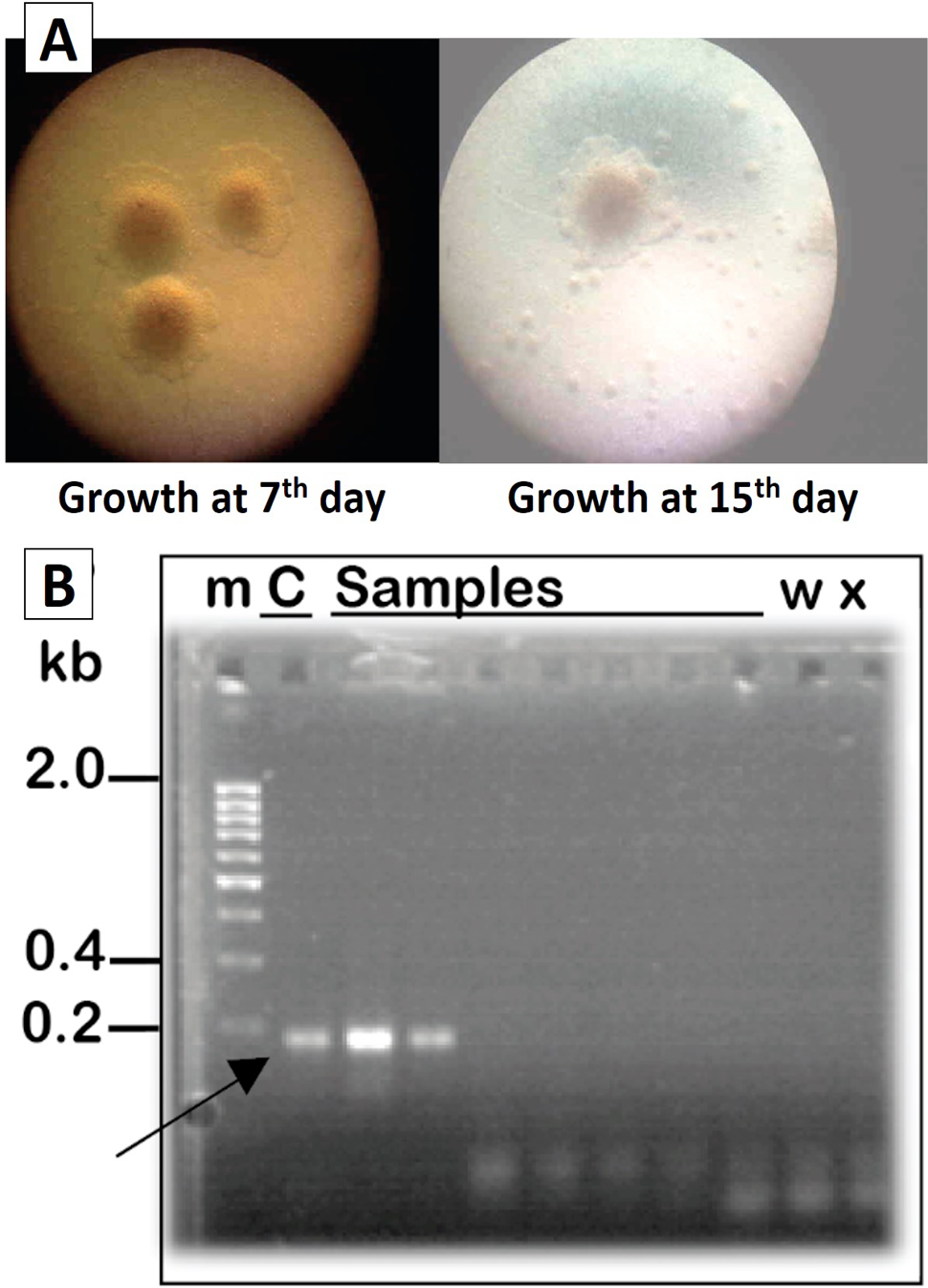
Detection and isolation of M. gallisepticum. Colonial morphology of M. gallisepticum is shown with left of 7 days post incubation, while, the right one was of 15 day post inoculation (c and d) Frequency (percentage) of detection of M. gallisepticum among isolates obtained after culturing swab samples on specific media (b) PCR-based detection of M. gallisepticum recovered from CRD suspected birds. Specie specific primers were used on the samples to amplify 185 bp long PCR product as shown by the arrow. The PCR amplicons are resolved on 1% agarose gel. The size was anticipated from a DNA ladder run in parallel. M, marker; C, positive control (DNA isolated from commercially available M. gallisepticum strain used for vaccination); w, water (genomic DNA was replaced with water for negative control); x, master mix and genomic DNA obtained from E. coli without water primers.