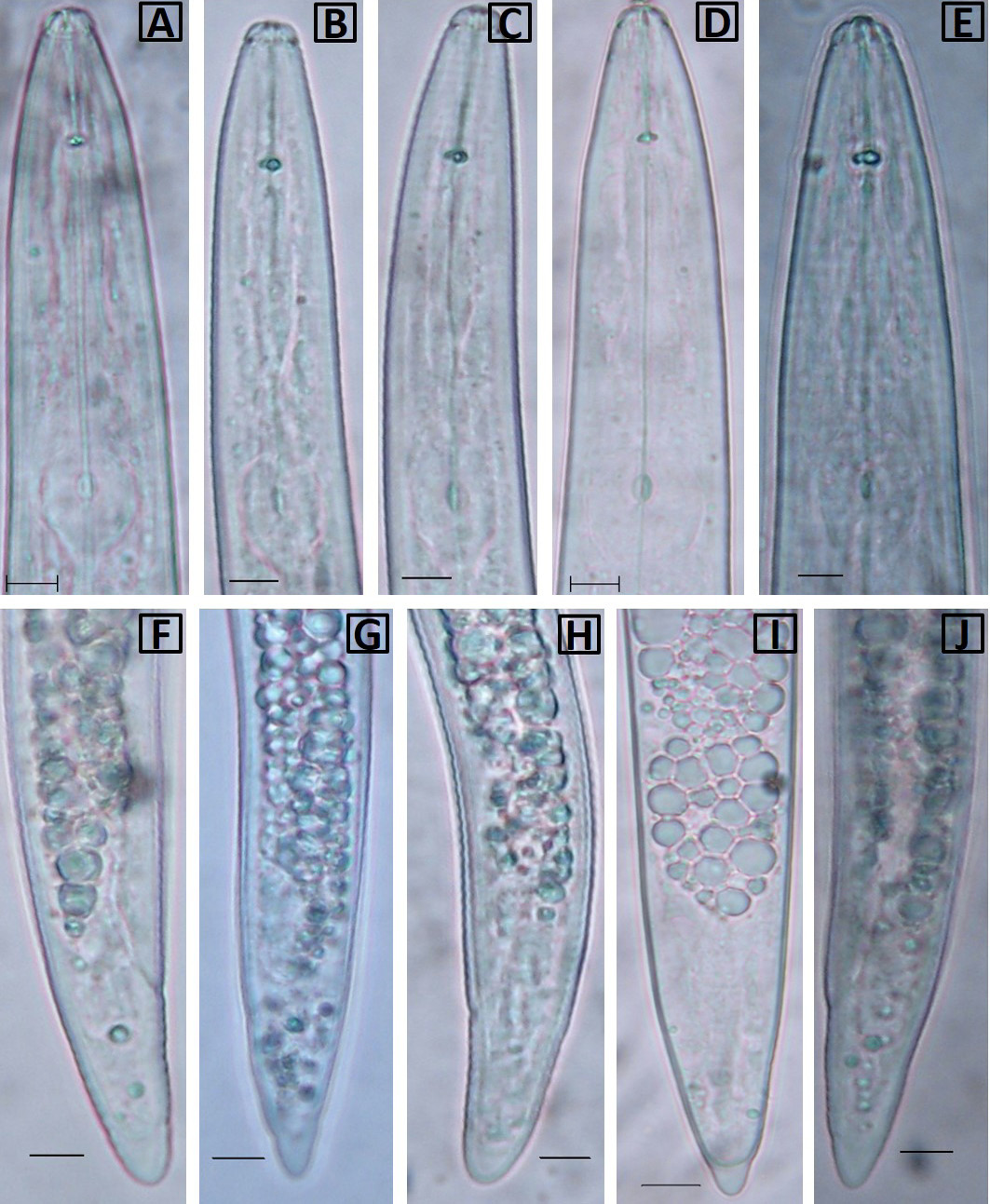
Variations in anterior end (A-E) and posterior end (F-J) of second stage juveniles. Scale –bar = 5 μm.