
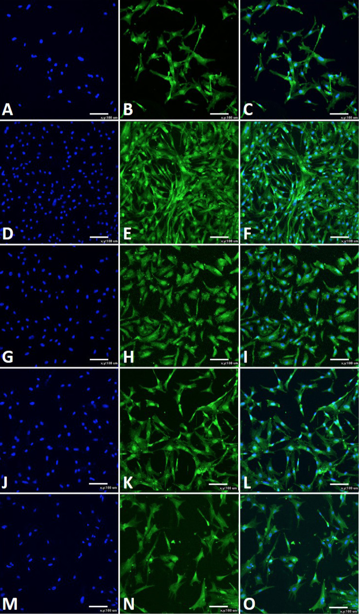
Fig. 4.
Immunofluorescence histochemical detection of CAFSCs. Cell surface markers Oct-4, CD105, nanog, CD73 and SSEA-4 are detected positive with immunofluorescence histochemistry on LSCM. A, D, G, J and M, nuclei is stained by DAPI; B, Oct-4+; E, CD105+; H, nanog+; K, CD73+; N, SSEA-4+; C, F, I, L and O, Merged.