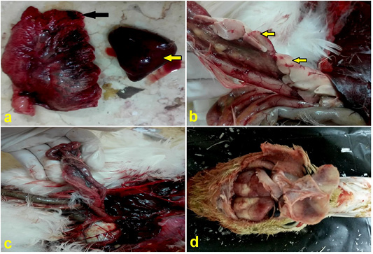
Figure 4
(a) Lung, duck showing congestion and edema (black arrow), spleen showing splenomegaly and congestion (yellow arrow). (b) Pancreas, duck showing necrotic areas (yellow arrows).) with hemorrhage (c) Duodenum, duck showing severe congestion of serosal vasculatures. (d) Brain, duck showing congestion.