
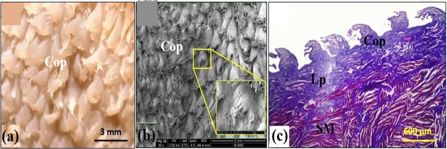
Fig. 3.
Light microscopical (a), scanning electron microscopic (b), and histological (Masson’s trichrome staining) structure of the lingual body in bear (c). Cop, conical papillae; Lp, lamina propria; SM, striated muscles. Yellow rectangle shows higher magnification of a conical papilla.