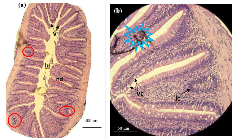
Figure 10:
Histology section of fore gut of catla infested with A. japonicus. a) Normal architecture of villi (v) with numerous gut associate lymphoid tissues in mucosal tissues (red circle) (10×); b) Emptied mucous cells (mc), aggregated lymphocyte cells (lc) (40×) =artifacts.