
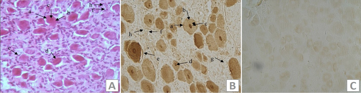
Fig. 1.
Immunohistochemical analysis of IL-18 Rα in IMG. A, the HE stains of the IMG of female goat; B, the immunohistochemical stains of IL-18Rα in the IMG; C, the blank control. a, membrane of IMG neuron; b, cytoplasm of the IMG neuron; c, nucleus of the IMC neuron; d, nucleoli of the IMC neuron; e, sertoli cells; f, blood vessels; g, schwann cell; h, endothelial cell.