
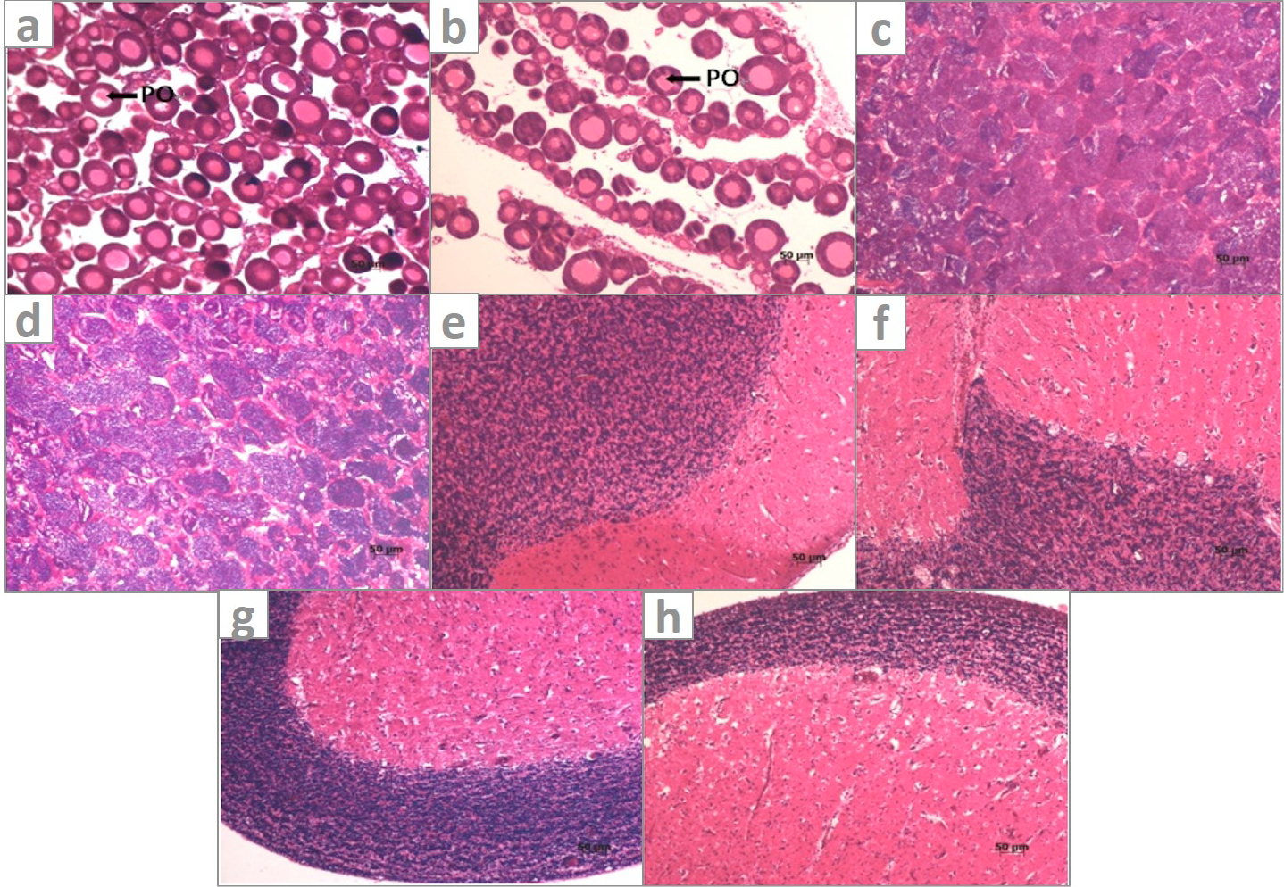
Fig 1
Histological structure of the gonads and brains of juvenile carp from control and the groups exposed to LET for 15d. a, Ovary from control groups showing abundance of large, closely arranged, uniformly developed primary oocytes; b, Ovary from LET(625μg/L)-treated groups showing increased prevalence of smaller early differentiated oocytes and enlarged ovarian plate space; c, Testis from control groups; d, from LET(625μg/L)-treated groups showing increased spermatogonial cells; e, brain from control male; f, brain from LET(625μg/L)-treated male; g, brain from control female; h, brain from LET(625μg/L)-treated female. PO, primary oocytes.