
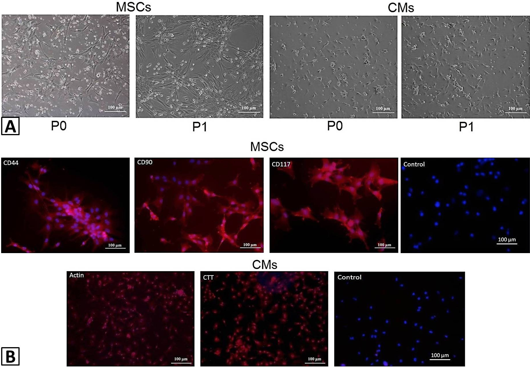
Fig. 1.
Morphology and charachterization of MSCs and CMs: (A) Morphology of rat bone marrow MSCs and rat CMs at passage 0 (P0) and passage 1. Images were taken under phase contrast at 10X magnification; (B) Characeterization of rat bone marrow MSCs and rat CMs. MSCs were treated with specific primary antibodies against CD44, CD90, and CD117. CMs were treated with specific primary antibodies against actin, and cardiac troponin T. Alexa fluor 546 secondary antibody was used for detection and DAPI was used for nuclei staining. Images were taken under fluorescence microscope.