
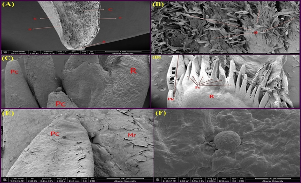
(A) SEM view of the dorsal cross section of the apex part of the tongue in the stud. (a) Stratified Squamous Epithelium (Keratinized), (K) Keratin, (A) Apex, (C) Corpus. (B) A higher magnification of the tongue apex with the stratified squamous epithelium cells, (a) Stratified Squamous Epithelium (Keratinized), (C) SEM view of the papillae of the radix part of the tongue (9-13 weeks). (R) Radix, (Plc) Papilla linguales caudales, (Pc) Papilla conicae. (D) SEM view of the papillae of the radix part of the tongue (stud). (R) Radix, (Plc) Papilla linguales caudales, (Pc) Papilla conicae. (E) SEM view of the papillae of the corpus-radix part of the tongue (9-13 weeks). (Pc) Papilla conicae, (Mr) Epithelial folds, (F) SEM view of the surface of the tongue body and papilla linguales (stud).