

Urine-Derived Mesenchymal Stromal Cells Alleviate Radiation-Induced TGF-β1 Production through the Inhibition of the NF-κB Signaling Pathway in MRC-5 Cells


Urine-Derived Mesenchymal Stromal Cells Alleviate Radiation-Induced TGF-β1 Production through the Inhibition of the NF-κB Signaling Pathway in MRC-5 Cells
Li-na Li1, Sheng Li2, Ping Gui3, Jing-hui Li1, Fen Ai4,* and Li-li Cai1,*

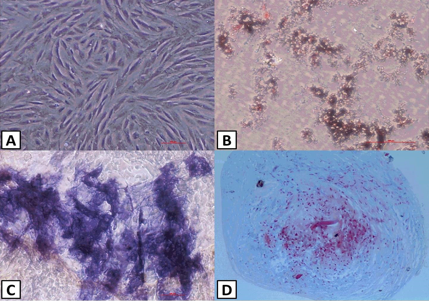
Identification of uMSCs. uMSCs exhibited a spindle- and fibroblast-like shape (A). B-D shows multipotential differentiation of uMSCs. uMSC differentiation into adipocytes, osteoblasts and chondroblasts, as shown by Oil Red O (B), alkaline phosphatase (C), and alcian blue (D) staining of in vitro differentiation cultures, respectively.
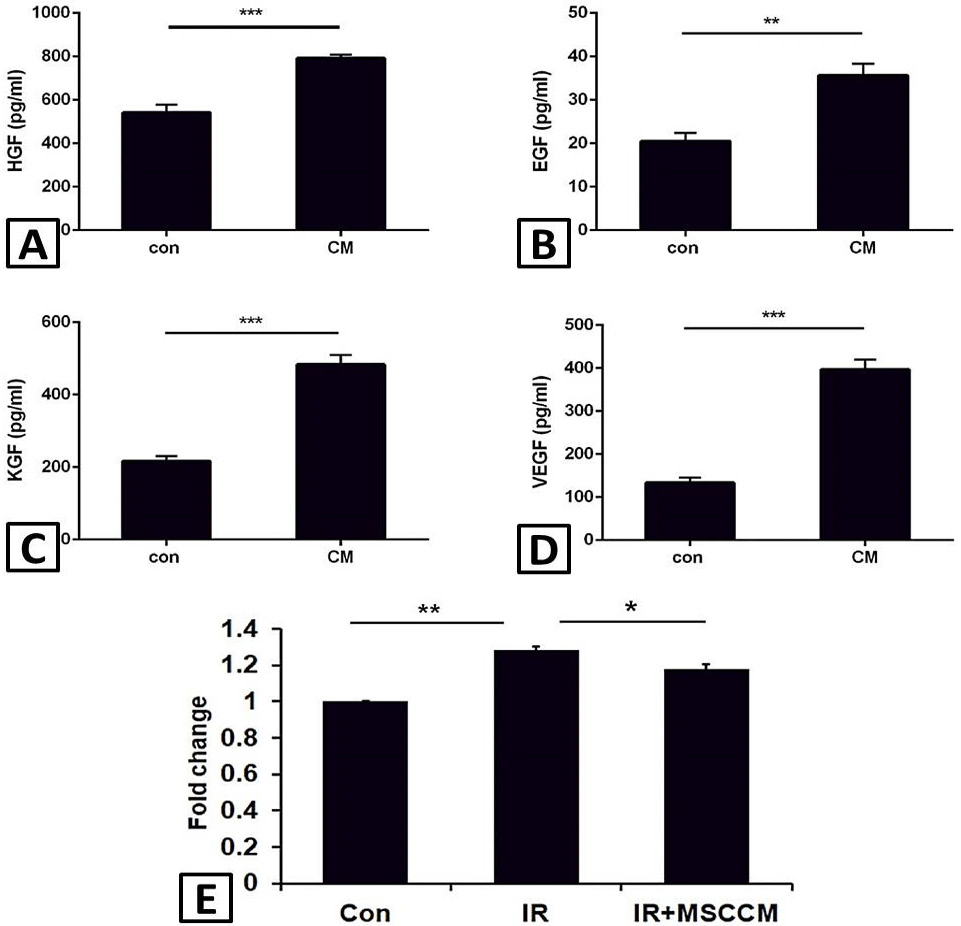
uMSCs secrete many types of protective cytokines and MSCCM inhibits the proliferation of MRC-5 cells. A-D, Expression of the cytokines of HGF, EGF, KGF and VEGF measured in the control media and the conditioned medium of uMSCs. E, cell viability analyzed by CCK8 assay. The results were compared among control cells (Con), single-irradiated MRC-5 cells (IR), and irradiation + MSCCM treated MRC-5 cells (IR+MSCCM). Data are expressed as the mean ± SD (n=5). *p< 0.05, **p< 0.01.
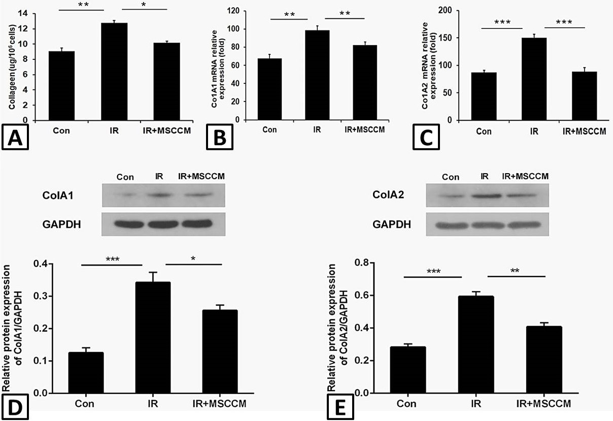
MSCCM reduces the collagen generation in MRC-5 cells. A, the collagen content was examined using Sircol soluble collagen assay in control cells (Con), single-irradiated MRC-5 cells (IR), and irradiation + MSCCM treated MRC-5 cells (IR+MSCCM). B-C, mRNA levels of Col1A1 (B) and Col1A2 (C) were detected by q-PCR. D-E, protein levels of Col1A1 (D) and Col1A2 (E) were detected by western blot. Data are expressed as the mean ± SD (n=5). *p< 0.05, ** p<0.01, ***p<0.001.
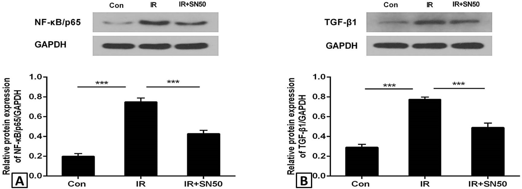
NF-κB inhibition could reduce irradiation induced upregulation of TGF-β1. A, protein level of NF-κB/p65 in different treatment groups detected by western blot. B, Protein level of TGF-β1 in different treatment groups detected by western blot. Data are expressed as the mean ± SD (n=5). *p< 0.05, **p<0.01, ***p<0.001.

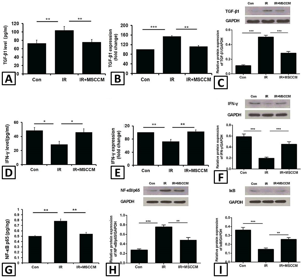
MSCCM inhibits the expression of TGF-β1 and prevents the NF-κB activation in MRC-5 cells after irradiation. A, D and G, TGF-β1, IFN-γ and NF-κB/p65 levels were determined by ELISA in lysate of MRC-5 cells in control cell group (Con), single-irradiated MRC-5 cell group (IR), and irradiation + MSCCM treated MRC-5 cell group (IR+MSCCM). B and E, the mRNA levels of TGF-β1 and IFN-γ were examined by q-PCR in different treatment groups. C, F, H and I, protein levels of TGF-β1, IFN-γ, NF-κB/p65 and IκB were detected by western blot. Data are expressed as the mean ± SD (n=5). **p<0.01, ***p<0.001.


