

Protective Effects of Intestinal Trefoil Factor against Endotoxin-Induced Injury of Intestinal Mucosal Epithelial Cells in Piglets


Protective Effects of Intestinal Trefoil Factor against Endotoxin-Induced Injury of Intestinal Mucosal Epithelial Cells in Piglets
Zhenyu Chang1, Hui Zhang1, Khalid Mehmood1,2, Mujeeb Ur Rehman1, Xiaodan Yuan1, Zhaoyang Li1, Fazul Nabi1, Xiaoxing Wu1, Xinxin Tian1, Xueting Liu1, Jingen Xu3 and Donghai Zhou1,*

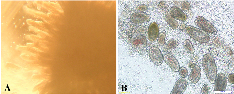
Intestinal villus, crypt units and lots of cells were detected by microscope, scale bars represent 100 μm.
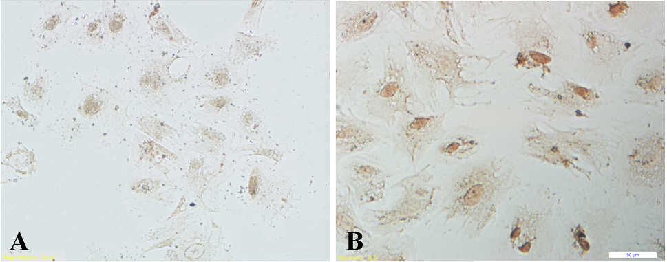
Immunodetection of cytokeratin in neonatal swine intestinal epithelia cells. Left: negative control, Right: epithelial cells assayed by Immunodetection of cytokeratin, scale bars represent 50 μm.
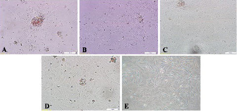
The culture of neonatal swine intestinal epithelial cell (a, after 48 h; b, after 72 h; c, after 96 h; d and e, overgrow; scale bars represent 100 μm).

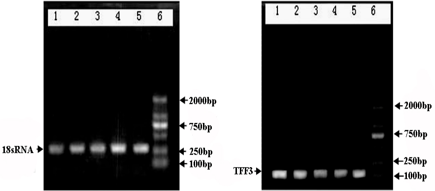
The results of PCR product 1% Agarose Gel Electrophoresis. Left, The PCR results of 18S rRNA. Right, The PCR results of TFF3. After total RNA reverse transcription and PCR, PCR products by 1% agarose gel electrophoresis, after the completion of the electrophoresis observe in the gel imaging system. The pictures shows the stripes clarity after PCR products by 1% agarose gel electrophoresis, reference gene 18S rRNA amplification, amplified fragment length of 275 bp, consistent with design. TFF3 target gene amplified fragment length of 174 bp, matches the designed length of the primers. Lane 1~4 respectively endotoxin final concentration of 0, 1, 10, 100 μg/mL group, lane 5 is TFF3 induced group, lane 6 is DL-2000 Marker. The results show that the internal gene and TFF3 gene PCR amplification effect is good, achieve the desired effect.

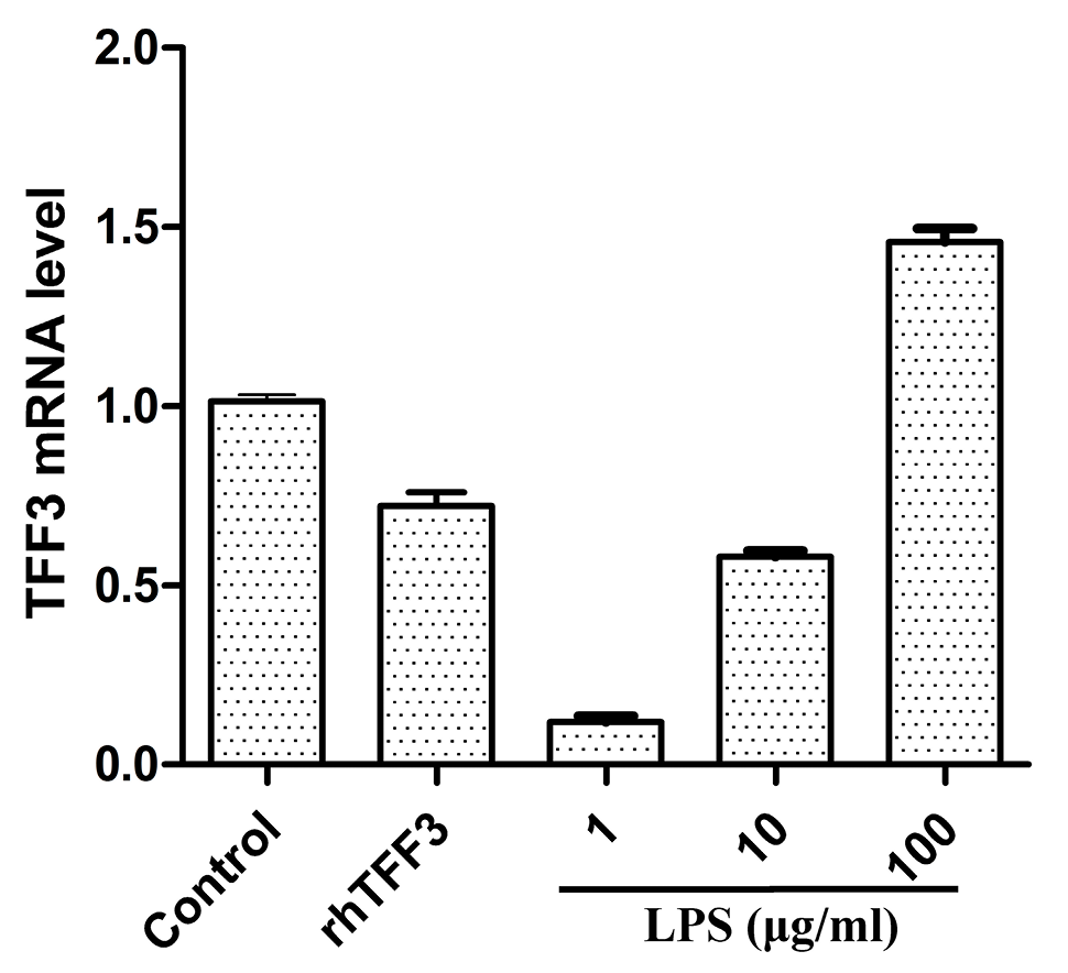
The expression of TFF3 genes in cells effect by LPS and rhTFF3. Cells were treated with a final concentration of, respectively; 1, 10, 100 μg/mL of endotoxin and 50 μg/mL of rhTFF3, the amount of TFF3 gene expression respectively 0.11, 0.58, 1.48, and 0.71 times of the control group, shows that concentration of 100 μg/mL of endotoxin plays an enhanced role in TFF3 gene expression in cells. With the concentration of 1 μg/mL, 10 μg/mL of endotoxin and 50 μg/mL rhTFF3, TFF3 gene expression in cells with different degree of inhibition.


