

Piceatannol Inhibits Akt Activation, Induces G2/M Phase Arrest and Mitochondrial Apoptosis and Augments Cisplatin Efficacy in U2OS Osteosarcoma Cells


Piceatannol Inhibits Akt Activation, Induces G2/M Phase Arrest and Mitochondrial Apoptosis and Augments Cisplatin Efficacy in U2OS Osteosarcoma Cells
Zijuan Li1,2, Rong Ma2, Muhammad Khan3*, Chenchen Liu2, Xiaolin Cui2 and Yongming Li1,2*
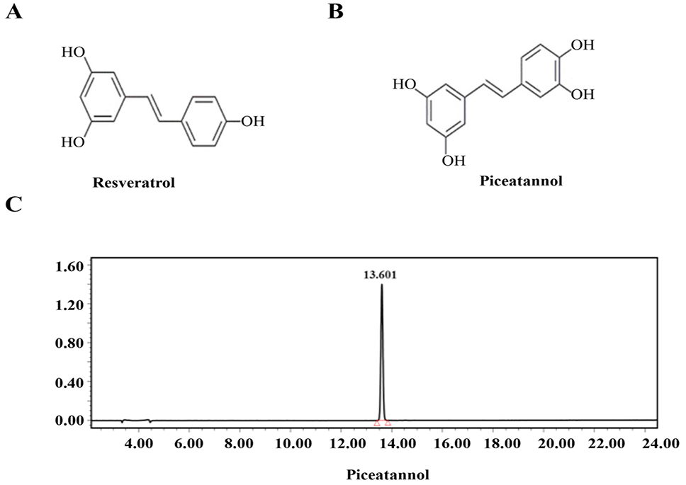
Chemical structure of medicine. (A) Chemical structure of Resveratrol. (B) Chemical structure of Piceatannol. (C) HPLC purity peak of piceatannol.

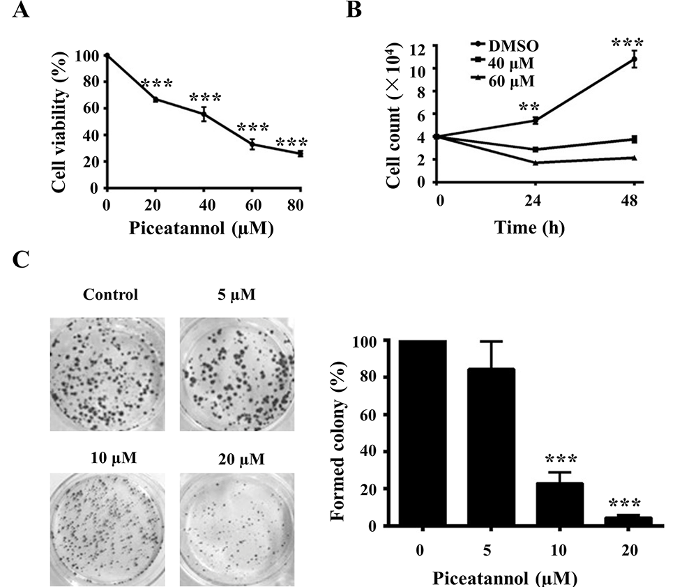
Piceatannol inhibits cell proliferation. (A) U2OS cells were treated with various concentrations of piceatannol for 72 h and cell proliferation was assessed using CCK-8 assay kit. (B) Cells were incubated with 0, 40 and 60μM for 24 and 48 h and finally 1X10⁴ cells were counted by flow cytometry. (C) Cell proliferation was measured by colony forming assay. The data is expressed as mean ± S.D. from three independent experiments. *p<0.05, **p<0.01, ***p< 0.001 significantly different compared with control.

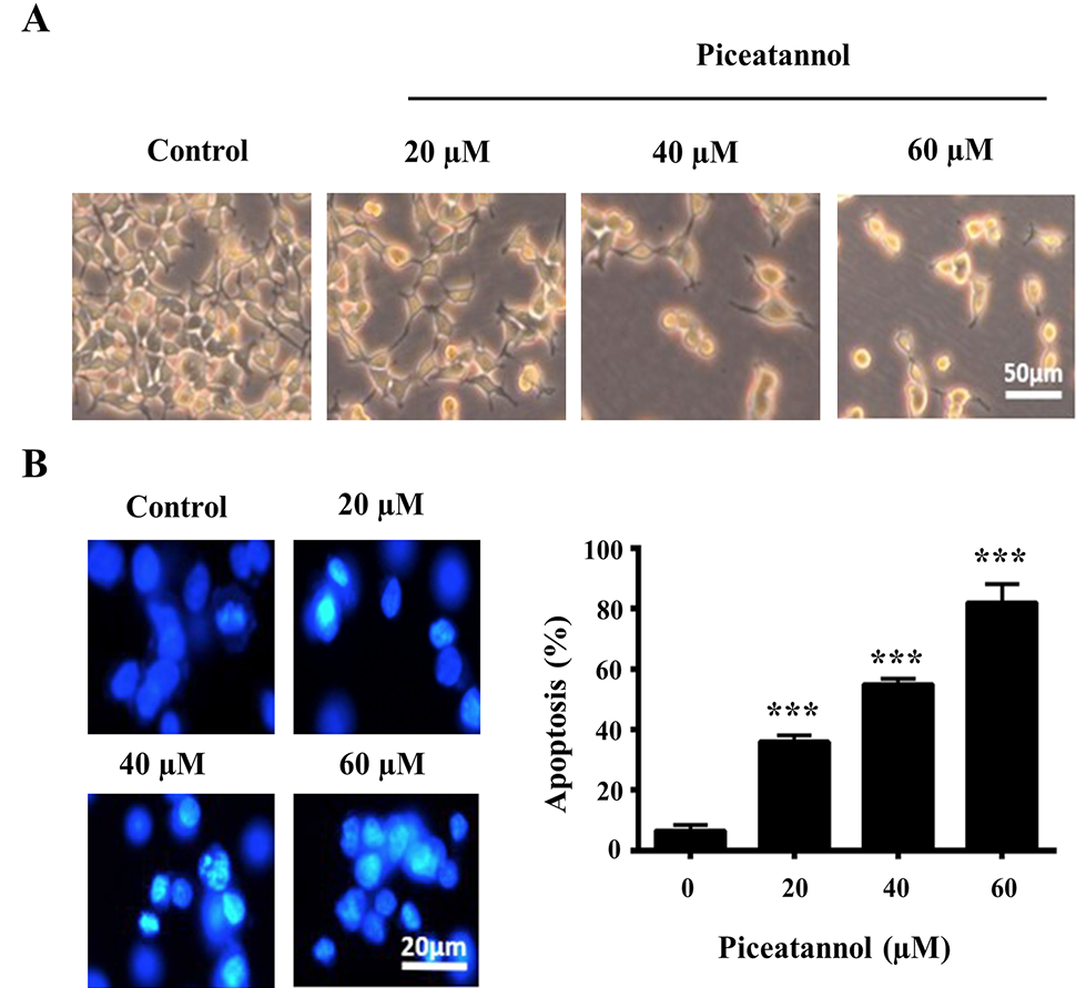
Piceatannol induces morphological changes and cell death in U2OS cells. (A) U2OS cells were incubated with various concentrations of piceatannol for 24 h and cellular morphology was observed under microscope. (B) U2OS cells were treated with different concentrations of Piceatannol for 24 h. Cells were stained with Hoechst 33258 and nuclear morphological changes were examined by fluorescence microscopy.
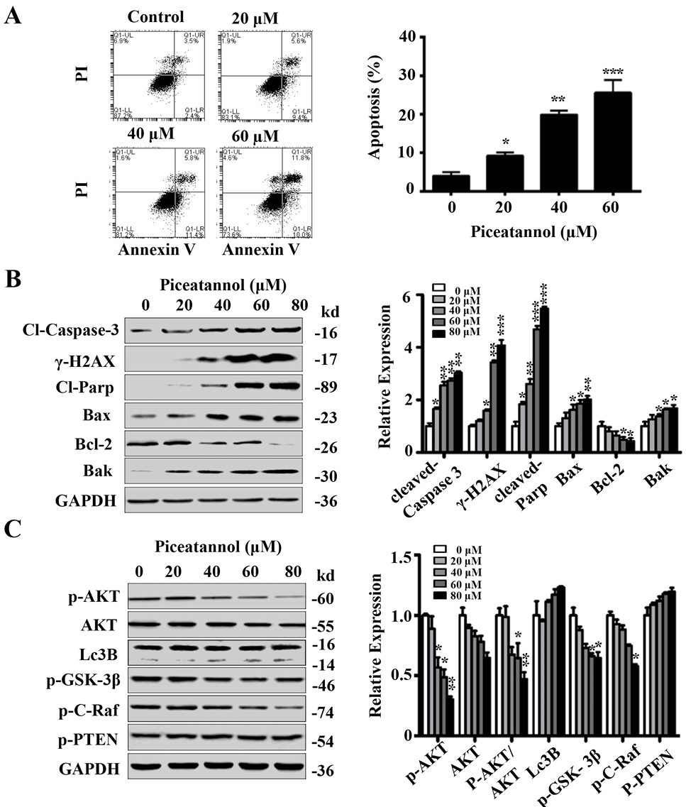
Piceatannol effectively induces apoptosis and suppresses PI3K/AKT pathway in U2OS cells. (A) Cells were stained with Annexin V/PI and apoptosis was determined by flow cytometry. (B) U2OS cells were treated with different concentrations of piceatannol for 24 h and expression of apoptosis regulating proteins was measured by Western blotting. (C) The inhibitory effect of piceatannol on PI3K/AKT pathway was assessed by Western blot. The bars in graph (B & C) represent the relative density of the bands normalized to GAPDH from three repeated experiments.

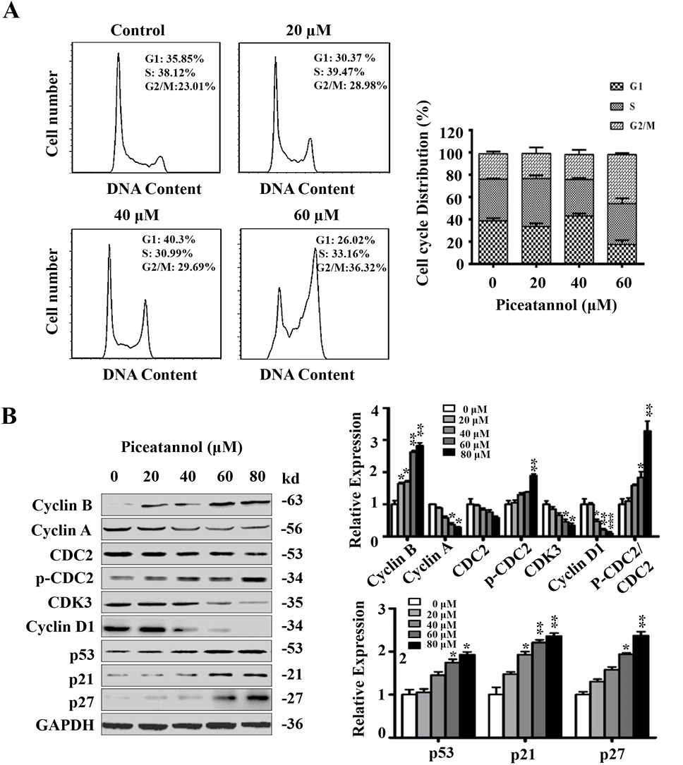
Piceatannol induces G2/M phase arrest in U2OS cells. (A) U2OS cells were treated with various concentrations of Piceatannol for 24 h. Flow cytometry was used to assess cell cycle profile. (B) U2OS cells were treated with Piceatannol for 24 h. The expression of cell cycle regulators was analyzed by Western blotting. The bars in graph represent the relative density of the bands normalized to GAPDH from three repeated experiments.
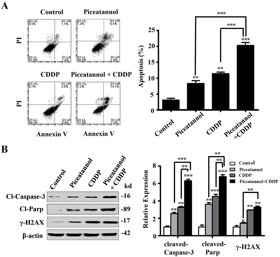
Combine treatment of piceatannol and CDDP induces apoptosis in U2OS cells with additive effect. (A) U2OS cell were treated with piceatannol (40μΜ), and CDDP (20μg/ml) either alone or in combination for 24 h. The apoptosis rates were measured by staining the cells with Annexin V/PI using flow cytometry. (B) The protein levels of cleaved-caspase-3, cleaved-PARP, and ɣH2AX were determined using Western blot in U2OS cell. The bars in graph represent the relative density of the bands normalized to β-actin from three repeated experiments.


