

Pathology of induced Velogenic Viscerotropic Newcastle Disease (VVND) in Japanese Quail and Myna


Pathology of induced Velogenic Viscerotropic Newcastle Disease (VVND) in Japanese Quail and Myna
Muhammad Faisal Ayoob1,2, Zaheer Ahmed Nizamani1, Asghar Ali Kamboh3, Mansoor Ayoob4, Waseem Ali Vistro3 and Abdul Sattar Baloch3*

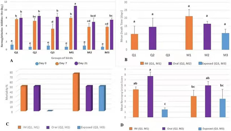
A, Haemagglutination inhibition titer mean values of serum sample(s) taken from experimental bird species (Japanese quail and myna) on Day 0, 7 and 21 of the experiment showing higher HI titers on day 21 as compared to day 0 and 7.
B, Mean death time (days) was recorded in experimental bird species (Japanese quail and myna) and early mortality was seen in Japanese quails as compared to mynas post-inoculation of VVND virus.
C, Mortality percentage was calculated in experimental bird species (Japanese quail and myna) and higher mortality rate was recorded in myna (75% intramuscular group) as compared to Japanese quail. Different groups of myna showed susceptibility to VVND whereas no mortality was recorded in Japanese quail (contact exposed group).
D, Dead birds were examined for postmortem findings and lesions on major infected organs and were scored according to severity of lesions. The average lesions score of oral groups of both experimental bird species (Japanese quail and myna) were higher than intramuscular and contact-exposed groups.
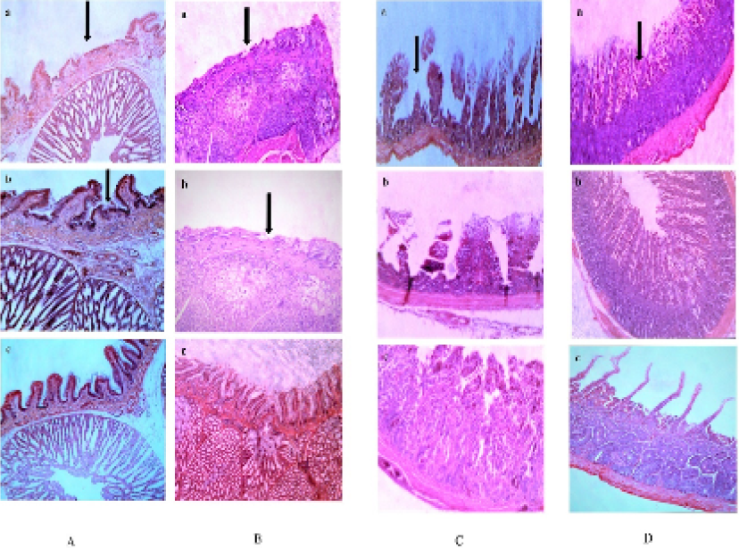
Histopathological lesions observed in proventriculus of Japanese quail (A) and myna (B) were mainly observed in the mucous membrane of the proventriculus. Extensive erosions in the epithelium, localized hemorrhages and congestion in the glandular region were observed. Glandular openings (villi) were swollen and had various degree of sloughed off mucosae, and irregular outline. Histopathological lesions observed in small intestine of Japanese quail (C) and myna (D) were mainly showed hyperemic and ulcerated microvilli. Surface desquamation was prominent and sloughing off the microvilli was also present.
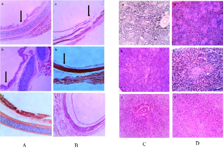
(A and B) Histopathological lesions observed in trachea of Japanese quail (A) and myna (B) were mainly discontinuation of surface epithelium. Loss of cilia of the lining epithelium and disturbance of coherence among various histologic layers were also noticeable. (C and D) Histopathological lesions observed in spleen of Japanese quail (C) and myna (D) were mainly enormous infiltration with mononuclear lymphocytes, especially in the area of red pulp. This region also showed hyperemic changes, petechial hemorrhages were also observed.


