

Osteometric and Radiographic Studies of Tarsal Bones in Adult Chinkara (Gazella bennettii)


Osteometric and Radiographic Studies of Tarsal Bones in Adult Chinkara (Gazella bennettii)
Salahud Din1,*, Saima Masood1, Hafsa Zaneb1, Habib Ur Rehman2, Saima Ashraf1, Imad Khan3, Muqader Shah4 and Syed Abdul Hadi1

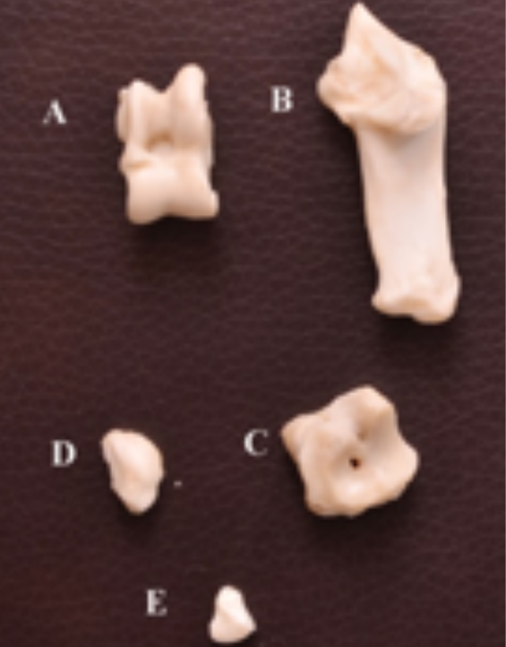
Tarsal bones of adult chinkara Talus (A); Calcanious bone (B); Central tarsal bone (C); fused 2nd and 3rd tarsal bone (D); first tarsal (E).

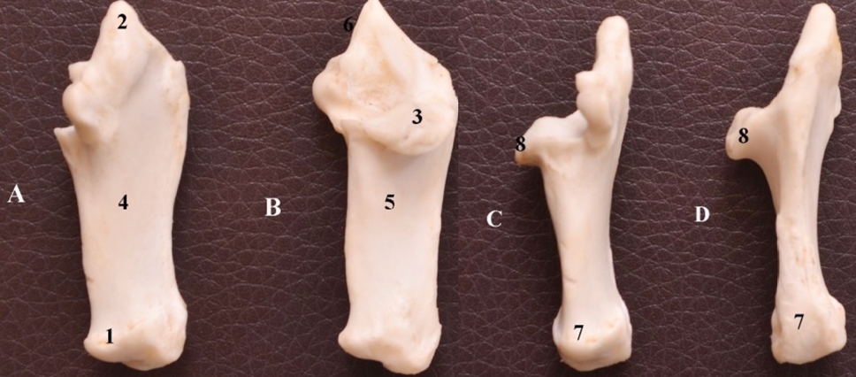
Different aspects (A, B, C, D) of adult chinkara fibular tarsal bone showing tuber calcis (1,7), facet for articulation with central and fourth fused tarsal (2), sustentaculum tali (3&8), lateral surface (4), medial surface.
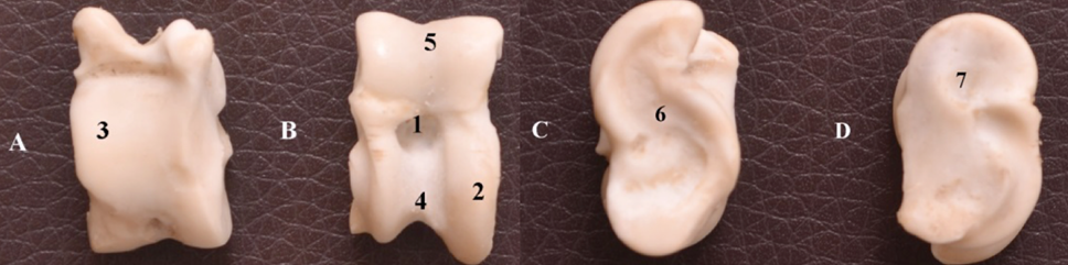
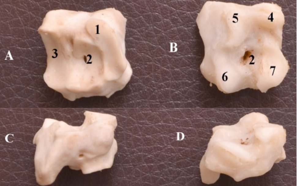
Surfaces of the tibial tarsal (A, B, C, D) of adult chinkara showing synovial fossa (1), condyles (2), planter surface bearing large oval facet (3), distal trochlea (4), proximal trochlea (5), medial surface (6), lateral surface (7).

Surfaces of the central and fourth fused tarsal bones showing proximal (A), ventral (B), medial (C), lateral surface (D), articular facets for calcanious bones. 1, foramen in the center of the bones more prominent on the ventral surface 2, articular facet for distal trochela of talus bone 3, articular facet for first, second and third fused tarsal (4,5,6 and 7).
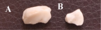
2nd and 3rd fused and first tar sal bone.

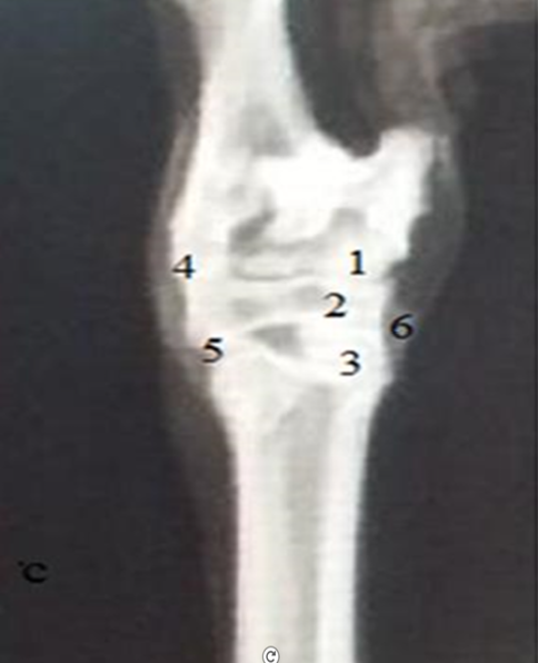
Dorso-palanter radiograph of the tarsal bones in adult chinkara proximal intertarsal (calcaneoquartal) joint. 1, distal inter tarsal joint; 2, tarsometatarsal joints; 3, articulation of calcanious and central and fourth tasral bone (on the lateral side of the hock); 4, articulation of central and fourth tasral bone and large metatarsal (on the lateral side of the hock); 5, superimposed first tarsal bone and 6, second tarsal bone.

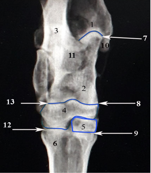
Palanto- dorsal radiograph of the tarsal bones in adult chinkara showing tibia (1), calcanious bone (2), talus bone (3), central tarsal bone (4), second and third fused Tarsal Bone (5), large metatarsal bone (6), tarsocrural joinfirt (7), proximal intertarsal (calcaneoquartal) joint (8), tarsometatarsal joints (9), Malleolus (10), sustenticulam tali (11), articulation of central and fourth tasral bone and large metacarpal (12), articulation of calcanious and central and fourth tasral bone (on the lateral side of the hock (13).

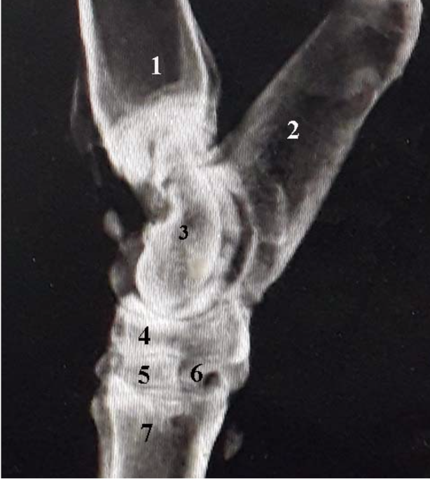
Mediolateral radiograph of the tarsal bones in adult chinkara showing tibia (1), calcanious bone (2), talus bone (3), central tarsal bone (4), second and third fused Tarsal Bone (5), first tarsal bone (6), large metatarsal bone (7).


