

Morphology of Lingual Papillae of Bear: Light Microscopic and SEM Study


Morphology of Lingual Papillae of Bear: Light Microscopic and SEM Study
Essam H. Ibrahim1,2,3, Attalla F. El-kott1,4, Ali Alshehri1, Mona Kilany2,5, Reza Yavari6, Salahud Din7* and Diaa Massoud8,9

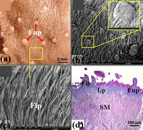
Light microscopical (a), scanning electron microscopic structure (b, c), and structure of the dorsal surface of the tongue of bear (d). Fup, fungiform papillae; red arrows, taste pores; Fip, filiform papillae; Lp, lamina propria; SM, striated muscles; yellow rectangles magnify the fungiform and filiform papillae in (b) and (c), respectively.
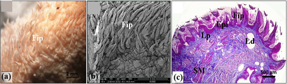
Light microscopical (a), scanning electron microscopic (b), and histological (Masson’s trichrome staining) of the lateral border of the tongue of bear (c). Lp, lamina propria; Ld, lipid droplets. For other abbreviations, see Fig. 1.

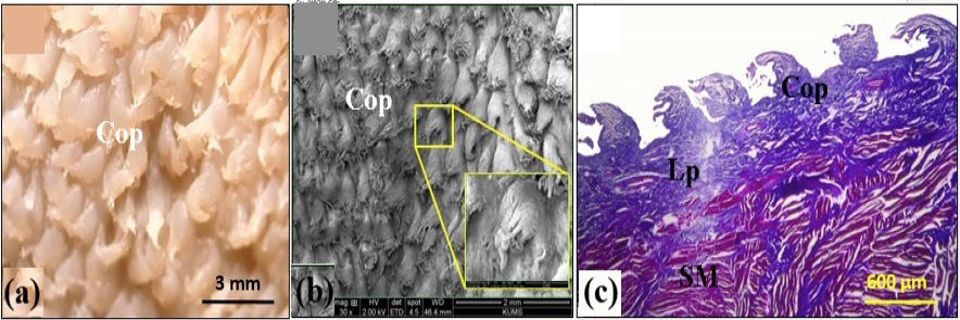
Light microscopical (a), scanning electron microscopic (b), and histological (Masson’s trichrome staining) structure of the lingual body in bear (c). Cop, conical papillae; Lp, lamina propria; SM, striated muscles. Yellow rectangle shows higher magnification of a conical papilla.

Light microscopical (a), scanning electron microscopic (b), and histological (Masson’s trichrome staining) structure of the lingual root in bear. Valp, vallate papillae; Fip, filiform papillae (c,d,e); Ap, annular pad; Gg, gastatory groove; Sg, salivary gland. For other abbreviations see Figure 1, 2 and 3. Red and black rectangles in (b) and (e) show higher magnification of a vallate papilla and a salivary gland accini.


