

Morphological and Molecular Characterization of Polydora websteri (Annelida: Spionidae), with Remarks on Relationship of Adult Worms and Larvae using Mitochondrial COI Gene as a Molecular Marker


Morphological and Molecular Characterization of Polydora websteri (Annelida: Spionidae), with Remarks on Relationship of Adult Worms and Larvae using Mitochondrial COI Gene as a Molecular Marker
Lingtong Ye1, Chao Cao1,2, Bin Tang1,2, Tuo Yao1, Ruixuan Wang1 and Jiangyong Wang1*
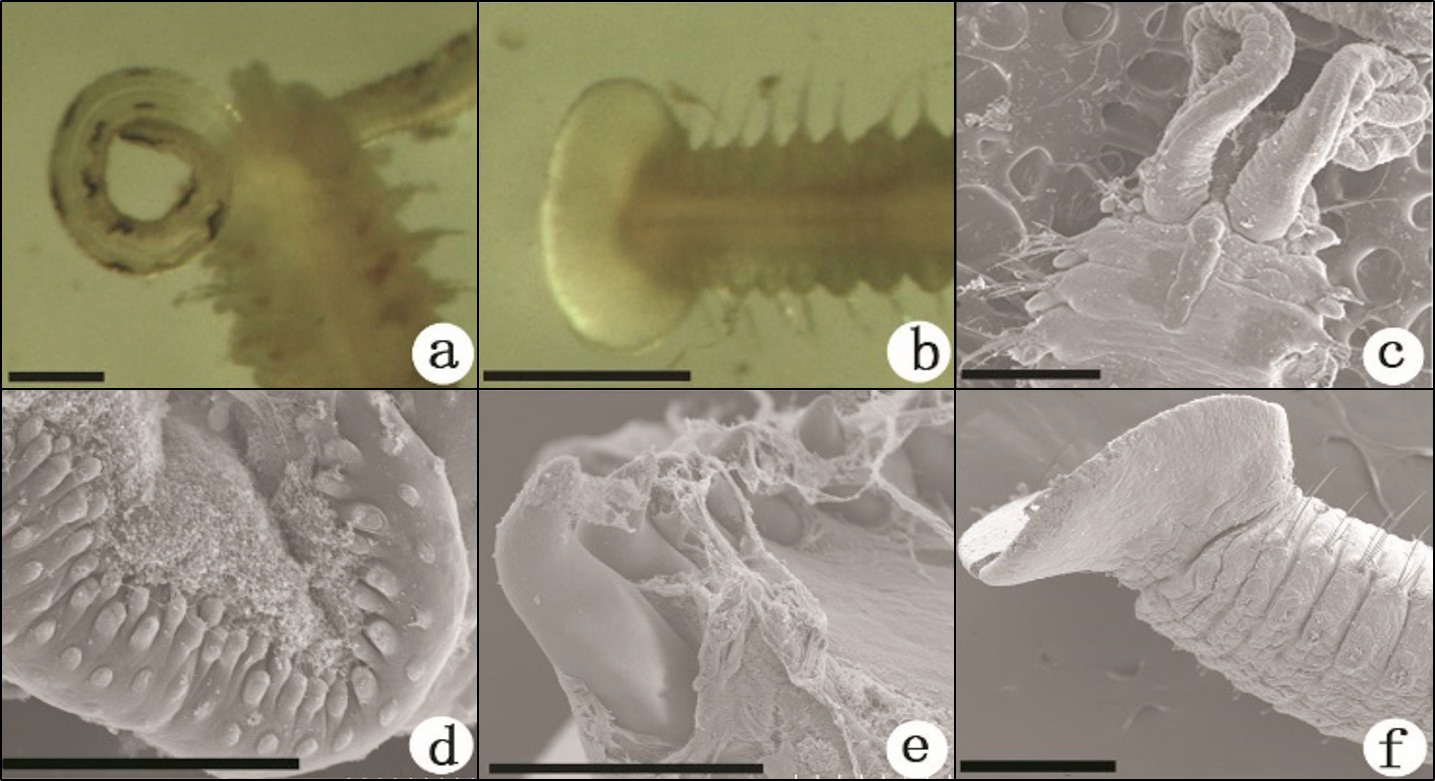
Fig 1
Adult worms of Polydora websteri. a–b, Light photographs; c–f, SEM images: a, Anterior end, dorsal view, showing non-continuous bar-like pigmentation line along the groove edges of palps; b, Posterior end, ventral view, showing cup-shaped pygidium; c, Anterior chaetigers with the caruncle extending back to posterior end of chaetiger 3; d, Margin of palps on which rod-like papillae are scattered; e, Heavy spines of chaetiger 5; f, Posterior chaetigers with cup-shaped pygidium. Scale bar = 50 µm.

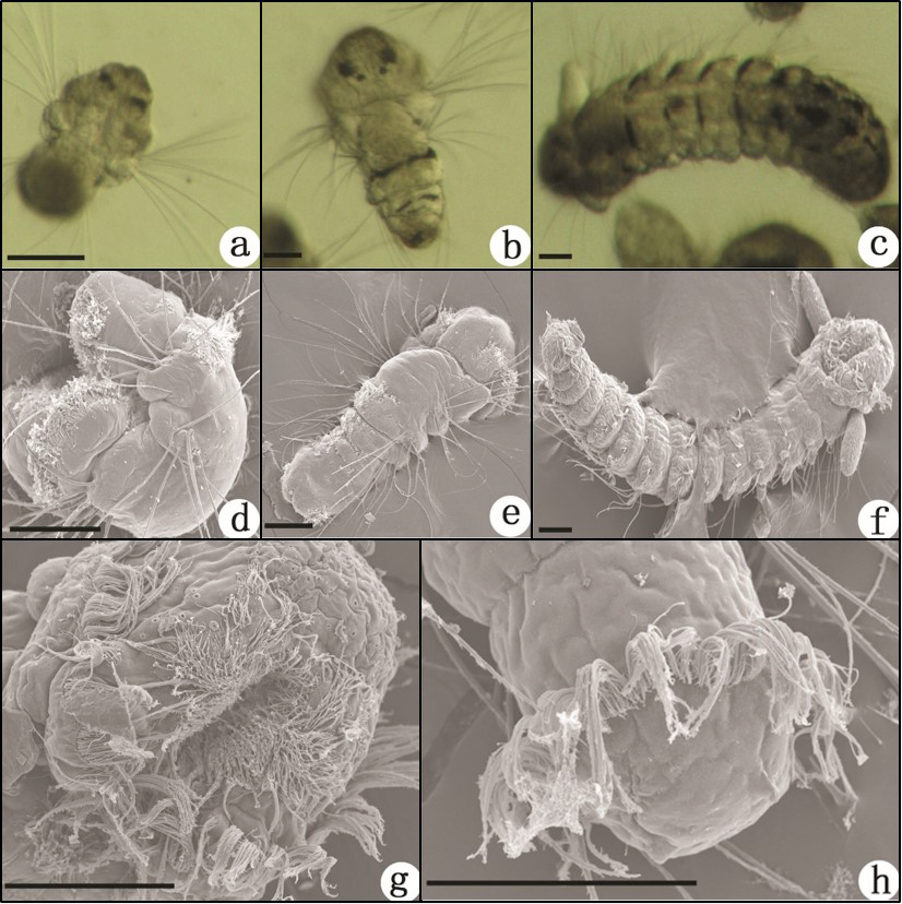
Fig 2
Larvae of Polydora websteri. a–c, Light photographs; d–h, SEM images: a, Three-chaetiger larva, dorsal view, showing two pigmented eyespots on the dorsal surface of the head; b, Five-chaetiger larva, dorsal view, two rows of pigment scattered across the dorsal surface of each chaetiger; c, Thirteen-chaetiger larva, dorsal view; d, Three-chaetiger larva, lateral view, serrated chaetae present on each side of the chaetigers; e, Five-chaetiger larva, dorsal view, the prototroch with a band of fine cilia, encircling the head, with the exception of the dorsal part; f, Thirteen-chaetiger larva, ventral view, a pair of round palps present on both sides of the head; g, Ventral view of the head, showing the vestibule with bundles of cilia; h, Dorsal view of posterior end, the telotroch with a circle of cilia. Scale bars = 50 µm.
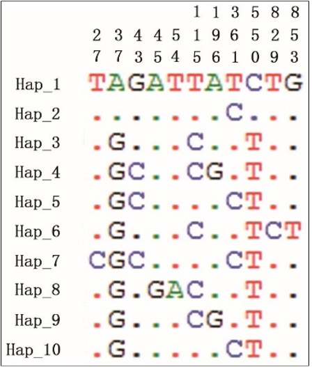
Fig 3
Location of mutative nucleotide acids in 10 haplotypes of mitochondrial COI sequences from Polydora websteri. Hap_1—Hap_10 represent the names of 10 haplotypes. Numbers on the top represent locations of mutative nucleotide acids in the 10 haplotypes. Dots indicate the bases which are the same as Hap_1.

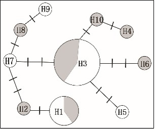
Fig 4
Mitochondrial haplotype network of Polydora websteri based on COI sequences. Network represents 10 haplotypes; 8 unique, 1 shared by 5 individuals and 1 shared by 12 individuals. Each perpendicular line represents one mutation step. The areas in grey represent the larval individuals, and the areas in white represent adult individuals. The areas of the circles are proportional to the number of samples of each haplotype.

April 2017
Vol. 49, Iss. 2, Pages 425-759

