

Internalization and Nuclear Localization of Porcine Growth Hormone in Porcine Hepatocytes


Internalization and Nuclear Localization of Porcine Growth Hormone in Porcine Hepatocytes
Pan Hong, Ruonan Li, Yumeng Li, Hainan Lan* and Xin Zheng*

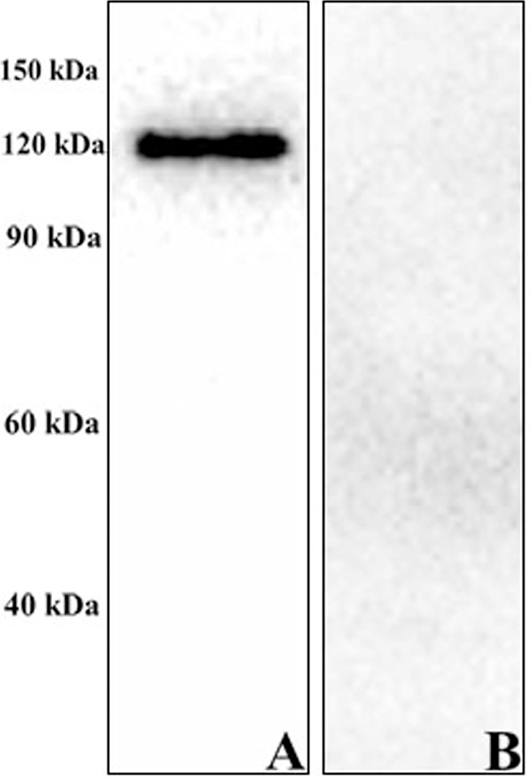
Western blot analysis to detect the expression of pGHR in porcine hepatocytes. Porcine hepatocytes were collected and lysed using cell lysis buffer, and the cell extracts were analysed by Western blotting using an anti-growth hormone receptor antibody (A) or control antibody (Rabbit IgG) (B). The figures represent at least three independent experiments.

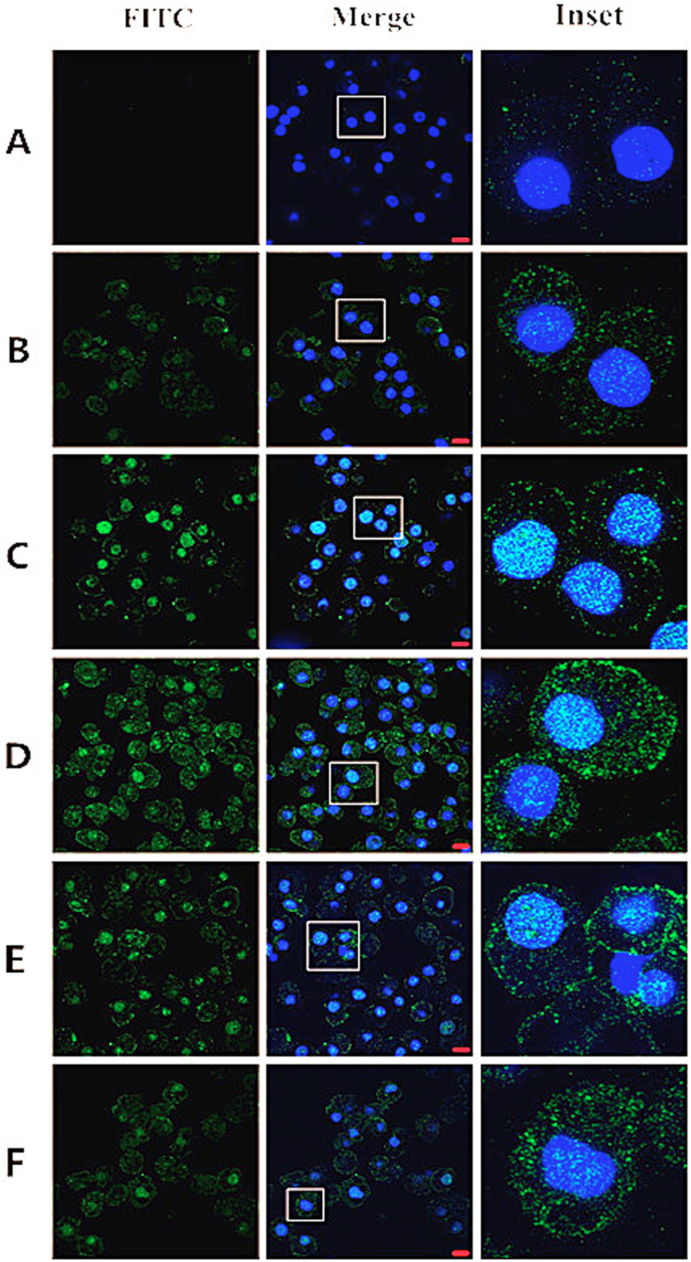
CLSM analysis to observe the internalization and nuclear localization of pGH in porcine hepatocytes. Porcine hepatocytes were serum starved, washed and incubated with FITC-pGH (100 ng/mL) for 0, 15, 30, 60, 75 and 90 min and subsequently fixed with 4% paraformaldehyde and stained with Hoechst 33258 (nucleus) (A-F). The cell samples were visualized by confocal laser scanning microscopy. Bar = 10 μm. The images represent at least three independent experiments.
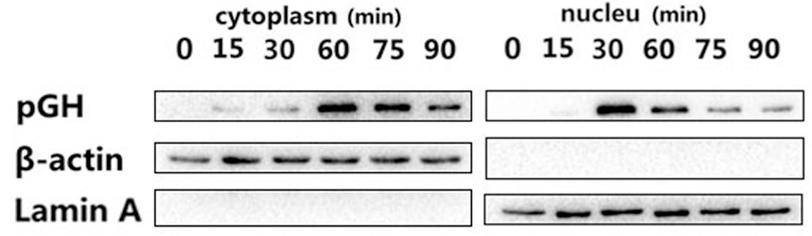
Western blot analysis to detect the internalization of pGH in porcine hepatocytes. Porcine hepatocytes were incubated with FITC-pGH (100 ng/mL) for different durations, and the cytoplasmic and nuclear extracts were subjected and analyzed by Western blotting using 1A3 (the mouse monoclonal antibody to pGH was generated in our laboratory). Anti-β-actin and anti-lamin A antibodies were used as markers for the cytoplasmic and nuclear extracts, respectively. The experiment was performed at least three times.
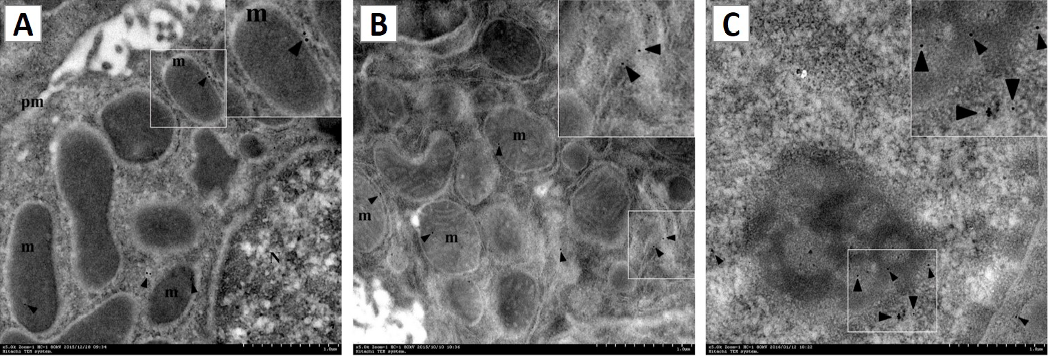
Subcellular localization of pGH. Immunoelectron microscopy samples were prepared as described in the “Materials and methods” section. Gold grains were localized to the cell membrane (pm), mitochondria (m) and cell nucleus (N) (A-C). Bar = 1μm. The micrograph represents at least three independent experiments.


