

In Silico Prediction and Evaluation of E. coli Expressed Recombinant HA Protein of Avian Influenza Virus


In Silico Prediction and Evaluation of E. coli Expressed Recombinant HA Protein of Avian Influenza Virus
Sana Shakoor, Tahir Rehman Samiullah, Naila Shahid, Abdul Qayyum Rao*, Aneela Yasmeen, Sana Tahir, Ayesha Latif, Saira Azam, Ahmad Ali Shahid and Tayyab Husnain
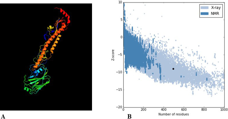
Prediction of immunogenic HA protein epitopes via bioinformatics tools. A, Image coloured by rainbow N → C terminus model dimensions (Å):X:46.119 Y:134.537 Z:45.524; B, Plot of residue scores.

Graphical representation of the occurrence frequency of HA protein residues based on individual score (Y-axis) and residue position (X-axis).

JSmol-rendered 3D structures of continuous antigenic epitopes of the HA protein along with their PI values, as predicted by ElliPro.

Confirmation of cloning of HA gene in pET30a expression vector and E. coli BL21 (D3). A, Confirmation of successful digestion of TA vector containing HA gene and pET30a vector; B, Confirmation of HA gene-pET30a Ligation through restriction digestions; Lane 1-12, Restriction digestion of pET30a clones, Lane L, 1Kb DNA ladder; C, Confirmation of HA gene ligated in pET30a through PCR through PCR; Lane 1-3, Amplification of 1663bp of HA gene in pET30a; D, Confirmation of presence of HA gene ligated in pET30a transformed into Rosetta by PCR; Lane 1-7, Amplification of 1683bp HA gene ligated in pET30a plasmid; Lane 8, -ve control; Lane L, 1 Kb DNA Ladder.

Animal study: Confirmation of HA protein through immune dot blotting assay. The appearance of dot on spotted position confirmed the specificity of anti-HA antibodies produced. A, Blood from the control group; B, Blood from the experimental group after first and second immunization.

ELISA assay. ELISA determined the titer of antibody produced in immunogenic mice, Diluted serum in 1/500 and 1/1500 effectively work as antibody as compared to 1:1000 dilution.


