
Haematological and Histopathological Analyses of Levofloxacin Induced Toxicity in Mammals


Haematological and Histopathological Analyses of Levofloxacin Induced Toxicity in Mammals
Chaman Ara1*, Asmatullah1, Sehrish Kanwa1, Asma Chaudhary2, Ayesha Siddiqua1
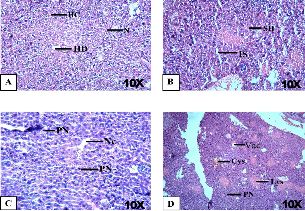
Histopathological examination of liver tissues of LFX exposed mice at 10X magnification. (A) Control mice showing normal structure i.e. HD: Hepatic duct; HC: Hepatocyte; N: nucleus. (B) Mice treated with 9.37 μg/g B.W. of LFX showing IS: increased sinusoidal spaces; SH: swollen hepatocytes. (C) Mice treated with 18.37 μg/g B.W. of LFX exhibiting Nc: necrosis; PN: pyknosis. (D) Mice treated with 37.50 μg/g B.W. of LFX showing showing defects i.e. Vac.: Vacuolation; PN: pyknosis; Lys: Lysed blood cells; Cys: cyst. H and E staining.

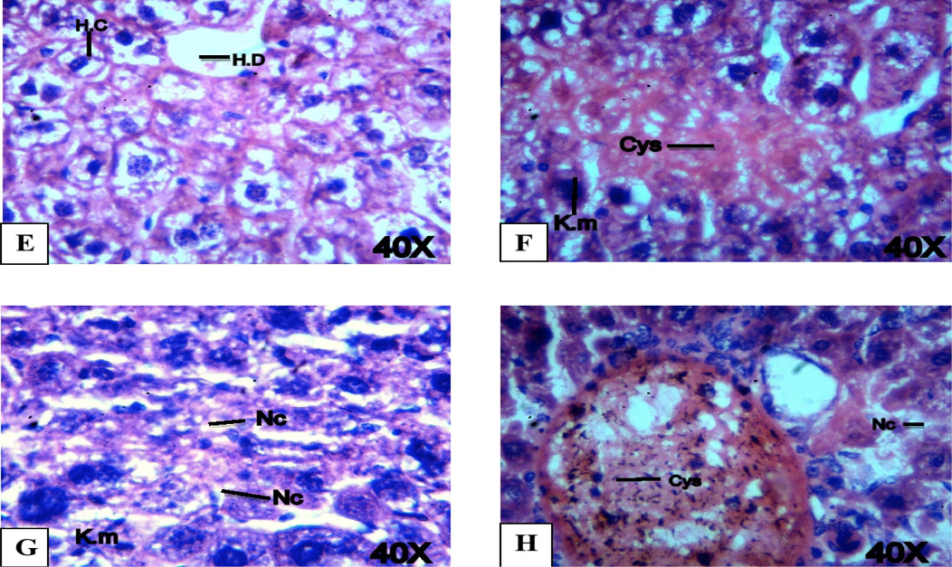
Histopathological examination of liver tissues of LFX exposed mice at 40X magnification. (A) Control mice showing normal structure i.e. HD: Hepatic duct; HC: Hepatocyte. (B) Mice treated with 9.37 μg/g B.W. of LFX showing Cys: cyst; K.m: karyomegaly. (C) Mice treated with 18.37 μg/g B.W. of LFX exhibiting Nc: necrosis; PN: pyknosis. (D) Mice treated with 37.50 μg/g B.W. of LFX showing Cys: cyst; Nc: necrosis’ H and E staining.

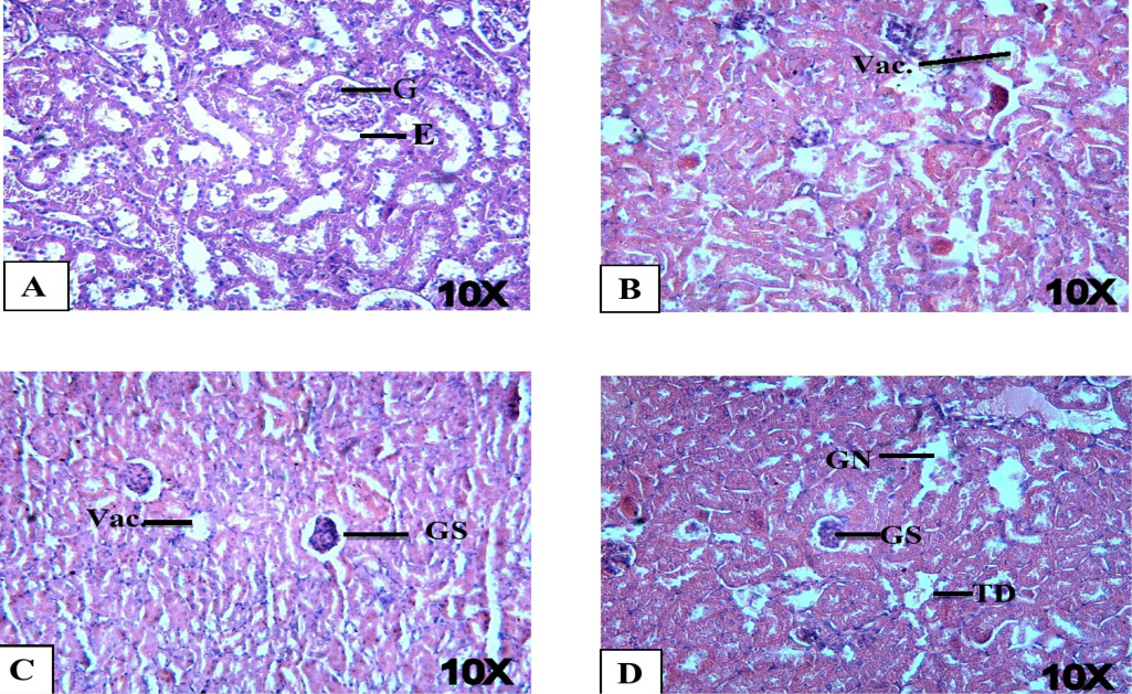
Histopathological examination of mice kidneys with LFX at 10X. (A) Control mice showing normal tissues i.e. G: glomerulus; E: epithelial lining. (B) Mice treated with 9.37 μg/g B.W. of LFX showing Vac.: vacoulation. (C) Mice treated with 18.37 μg/g B.W. of LFX showing GS: gomerulosclerosis; Vac.: vacoulation. (D) Mice treated with 37.50 μg/g B.W. of LFX showing GS: glomerulonephritis; GN: glomerulonephritis; TD: tubular degeneration. Stain H and E.

Histopathological examination of testes of LFX exposed mice at 10X magnification. (A) Control mice showing normal tissues i.e. SP: sperm; S.T.: semeniferous. (B) Mice treated with 9.37 μg/g B.W. of LFX showing Ex.F: exfoliation of spermatocytes; R.E: Rapture of epithelium. (C) Mice treated with 18.37 μg/g B.W. of LFX exhibiting A.S: spermia; Ex.F: exfoliation of spermatocytes; T.D: tuber degeneration. (D) Mice treated with 37.50 μg/g B.W. of LFX showing Ex.F: exfoliation of spermatocytes; Vac: vaculation; F.D: fat deposition, A.S: Aspermia. H and E staining.
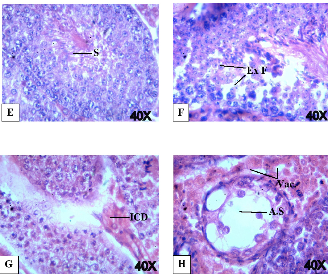
Histopathological examination of testes of LFX exposed mice at 40X magnification. (E) Control mice showing normal tissues i.e. SP: sperms. (F) Mice treated with 9.37 μg/g B.W. of LFX showing Ex.F: exfoliation of spermatocytes. (G) Mice treated with 18.37 μg/g B.W. of LFX exhibiting ICD: interstitial cell degeneration. (H) Mice treated with 37.50 μg/g B.W. of LFX showing Vac: vaculation; A.S: Aspermia. H and E staining.
