
Effect of Experimentally Induced Depression on the Adrenal Cortex of Adult Male Albino Rats and the Possible Ameliorative Role of Voluntary Exercise


Effect of Experimentally Induced Depression on the Adrenal Cortex of Adult Male Albino Rats and the Possible Ameliorative Role of Voluntary Exercise
Magda A. Eldomiaty1,2, Manal E. Elsawaf2, Soad S. Ali3, Heba El-Sayed Mostafa4,5*

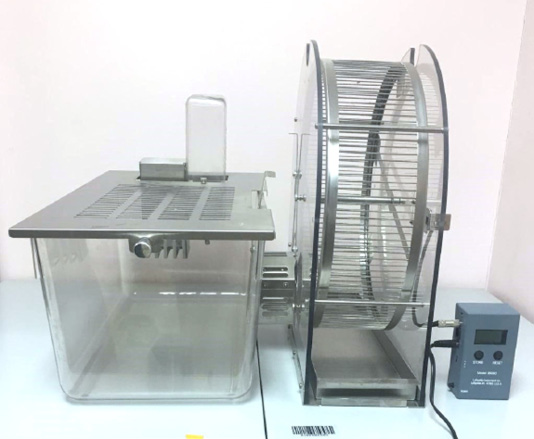
A cage equipped with voluntary rat running wheel with activity wheel counter.

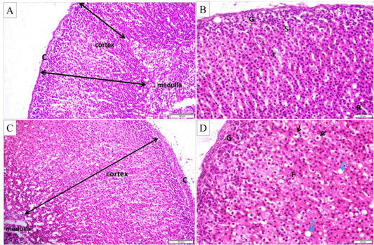
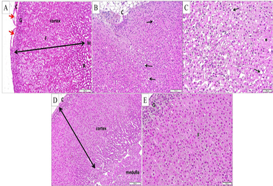
(A and B) Histological structure of adrenal gland in control group (H and E stain X200 and X400 respectively), showing the capsule (c), cortex and medulla. The cortex is formed of three zones; zona glomerulosa (G), fasciculata (F) and reticularis (R). Notice the blood sinusoids (s). (C and D) Histological structure of adrenal gland in the control exercise group (H and E stain X200 and X400, respectively), showing apparent increase of cortical thickness with hypertrophied cells (black arrows) and large cytoplasmic vacuoles (green arrows) in zona fasciculata.

(A, B and C) Adrenal gland from depression group (H and E stain A and B X200 and C X400, respectively), showing. (A) Cortical zones; glomerulosa (G), fasciculata (F) and reticularis (R) and the medulla (M), with apparent increase of cortical thickness and apparent intracapsular cavities (red arrows). B) Disorganization of cortical zones and fat deposition in the capsule (c). (C) Hypertrophied cells in zona fasciculata with large cytoplasmic vacuoles (black arrows). Some cells show absent nuclei (*). (D and E) Adrenal gland from the depression exercise group (H and E stain X200and X400 respectively) with restoration of the normal adrenal architecture and normal cellular organization in zona glomerulosa (G) and fasciculata (F).
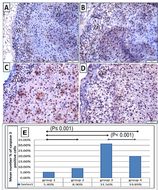
The immunohistochemical reaction of caspase 3 stained sections (X 400): (A) control group showing immune-positive cells with cytoplasmic reaction located mainly in zona granulosa and dispersed throughout zona fasciculata. (B) Control exercise group showing mild increase of the caspase3 positive cells in both zona glomerulosa and zona fasciculata. (C) Depression group showing extensive increase in the caspase3 positive cells in the zona granulosa, and aggregates of the immune-positive cells throughout the zona fasciculata (thick arrows). (D) Depression exercise group showing obvious reduction of immunoreactive cells in comparison to the depression group. The cells showed intense reaction and are dispersed in the zona glomerulosa and zona fasciculata (E) The mean numbers % of caspase 3 positive cells in different groups. Note: ZG, zona granulosa; and ZF, zona fasciculata.

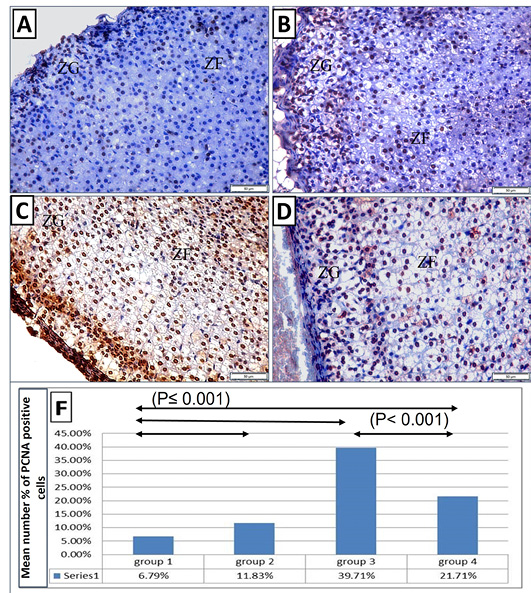
The immunohistochemical reaction of PCNA stained sections (X 400): (A) control group showing few sporadic immunoreactive PCNA positive cells with nuclear reaction in zona glomerulosa and zona fasciculata. (B) Control exercise group showing obvious increase of the PCNA positive cells in zona glomerulosa and zona fasciculata. (C) Depression group showing enormous increase of the PCNA positive cells with intense nuclear reaction in zona glomerulosa, at the junction between in zona glomerulosa and zona fasciculata and dispersed throughout zona fasciculata. (D) Depression exercise group showing less PCNA positive cells than those in the depression group, with the stained cells dispersed in zona glomerulosa and zona fasciculata. (E) The mean numbers % of PCNA positive cells in different groups.
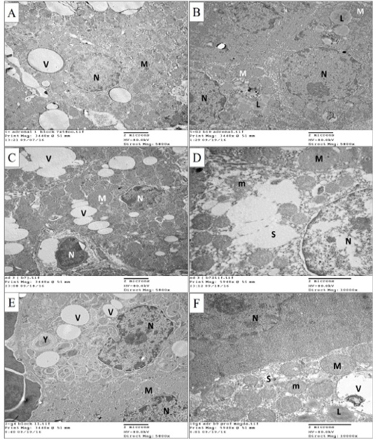
Electron microscopic pictures for the zona fasciculata cells of the adrenal cortex from different groups. (A) Cells of the control group showing rounded regular nucleus (N) with dispersed chromatin, multiple vesicular mitochondria (M) and vacuoles (V). (B) Cells of the control exercise group, showing rounded nuclei (N) with dispersed chromatin, multiple vesicular mitochondria (M). Notice the multiple vacuoles with lipid content (L). (C) Cells of the depression group showing shrunken irregular nuclei (N), swollen mitochondria (M) and multiple vacuoles of different sizes (V). (D) Cells of the depression group showing some swollen mitochondria (M), mitochondria with destructed cristae (m) and dilated vesicular smooth endoplasmic reticulum (S). (E) Cells of the depression exercise group showing normal sized nuclei (N) with irregular contour and multiple normal vesicular mitochondria (M). Few vacuoles (V) and lysosomes containing substance (Y) appear in the cytoplasm. (F) Cell from the depression exercise group showing normal nucleus (N), mitochondria with normal size and cristae (m) and nearly normal smooth endoplasmic reticulum (S). Few mitochondria appear dilated (M). Few vacuoles (V) appear with some showing lipid content (L). A, B, C and E x5800, and D and F x10000 - scale bar 2µ.

