

Digit and Sesamoid Bones of Adult Chinkara (Gazella bennettii) (Sykes, 1831) (Mammalia: Bovidae): Morphology and Osteometry


Digit and Sesamoid Bones of Adult Chinkara (Gazella bennettii) (Sykes, 1831) (Mammalia: Bovidae): Morphology and Osteometry
Salahud Din

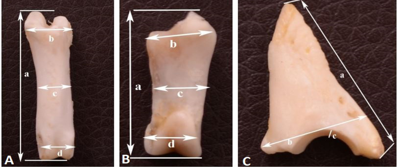
Dimensions of first (A), second (B) and third (C) phalanx of adult Chinkara. (A) greatest length (a) maximum breadth of proximal extremity (b) maximum breadth of shaft (c) maximum breadth of distal extremity (d). (B) greatest length (a), maximum breadth of proximal extremity (b), maximum breadth of shaft (c), maximum breadth of distal extremity (d). (C) greatest length (a), maximum breadth of proximal extremity (b), maximum breadth of articular surface.
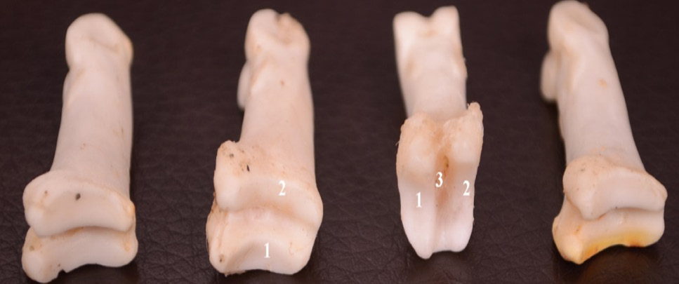
Proximal articular surface of the proximal phalanx of chinkara showing axial facet (1), abaxial facet (2) dorsopalmar groove (3).
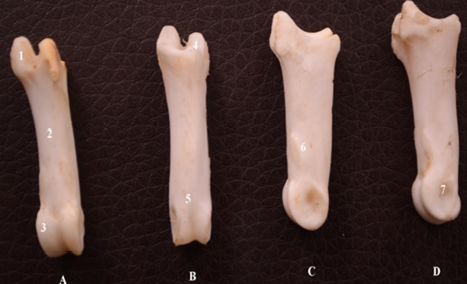
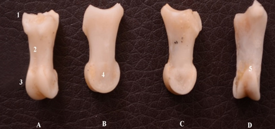
Different surfaces (palmar A, dorsal B, axial C, abaxial D) of the proximal phalanx of chinkara showing proximal extremity (1), shaft (2) distal extremity (3), dorsal side of articular surface (4), articular condyles (5), nodular elevation (6) axial depression (7).

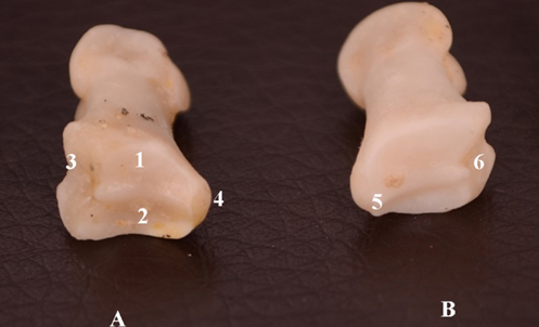
Different surfaces (A,B,C, D) of the middle phalanx of the forelimb of chinkara showing proximal extremity (1), shaft (2) distal extremity (3), axial depression (4), palmar surface of the articular condyles (5).
Proximal articular surface of the middle phalanx of the forelimb of chinkara showing axial facet (1), abaxial facet (2), palmar surface (3), dorsal surface (4), dorsopalmar ridge (5), palmarl depression (6).

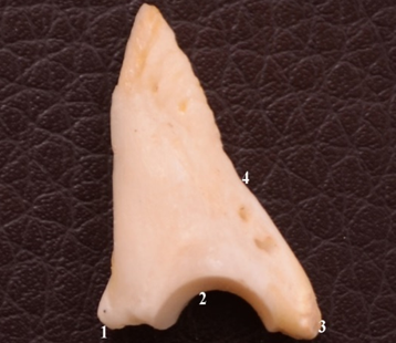
Third phalanx of the forelimb of chinkara showing extensor process (1), articular surface (2), ventral border (3, 4).
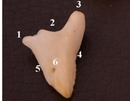
Third phalanx of the forelimb of chinkara showing extensor process (1), articular surface (2), ventral border (3, 4); dorsal border (5); caudal foramen (6).
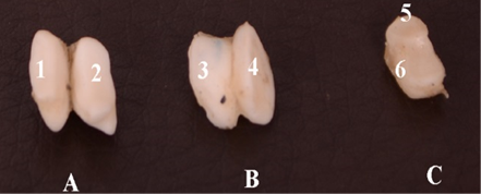
Dorsal (A) and palmar (B) view of the proximal sesamoids of chinkara showing axial sesamoid (1, 4); abaxial sesamoids (2, 3). Dorsal view of the distal sesamoids (C) showing articular surface (5, 6).


