
Comparative Prevallance and Pathological Changes on Camel Brucelosis at the Selected Slaughterhouses in Garissa County, Kenya


Comparative Prevallance and Pathological Changes on Camel Brucelosis at the Selected Slaughterhouses in Garissa County, Kenya
Abdirahman Barre*, Karanja D Njuguna, Bebora Lilly Caroline and George Chege Gitao
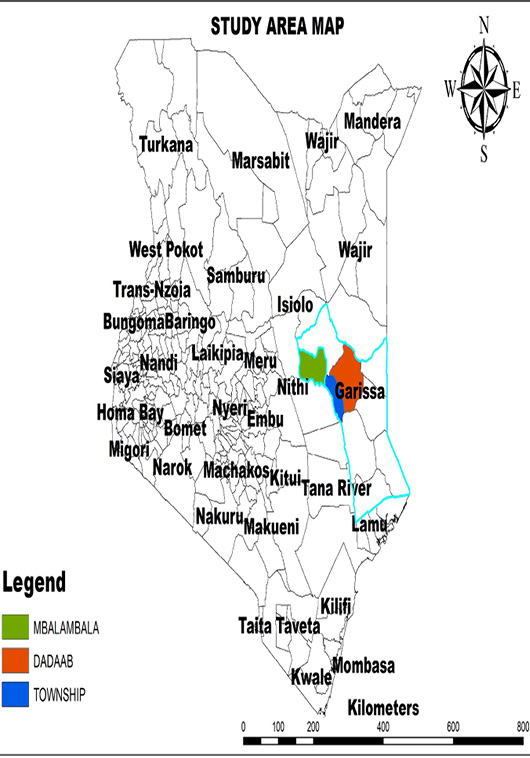
Map of Kenya showing that three sub counties and County of Garissa (Kenya Political Map, 2016; Kenya National Bureau of Statistics, 2017).
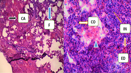

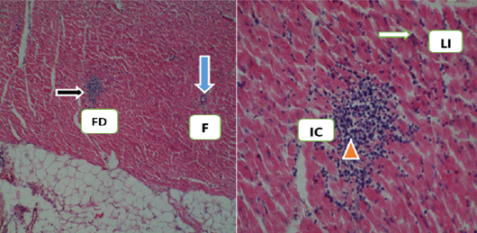
Those two (2) features were mad by Histological features of heart organ from zero -Positive tested camel obtained from Sample camel number (SC-GT- 30) were revealed that fatty degeneration (FD), lymphoblastic infiltration (LI) in cardiac muscles, slightly destructions of fibrous (F), macrophages, neutrophilic infiltrations in some areas and inflammatory cells (IC) (H/E × 40x and 100x).

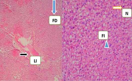
Histological section of condemned liver with tested seropositive obtained from sample camel Number (SC-24) was specified fatty degeneration (FD), Neutrophils (N), liver injuries (LI), diffuse fatty infiltration (FI) (H/E × 40x and 100x).

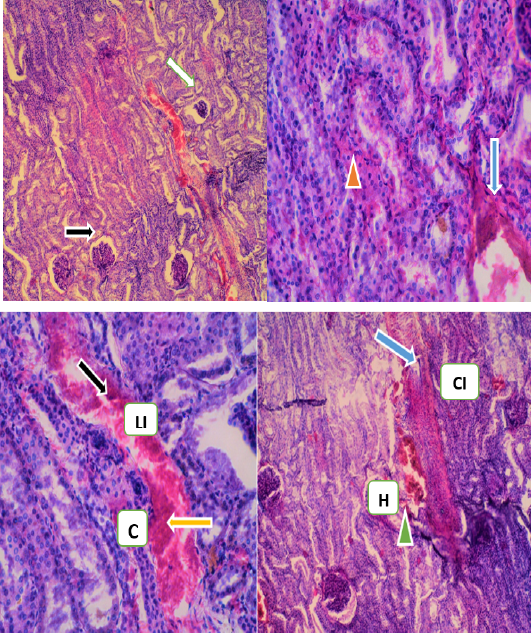
The histlogical features of kidney with tested seropositive obtained from sample camel numbe (SC-70) was pointed infiltration lymphoblastic cells (LI),congestions (C),cellulr infiltaraions (CI) and hemorhages (H) (H/E ×40, 400x and 100x).
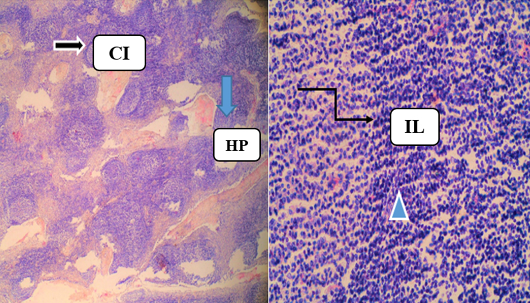
Histopathology of Lymph node for brucellosis-positive Camel case number (SC-12) and were viewed that the immunoblastic infiltration, Cellular infiltration (CI); hypoplasia of lymphocytes (HP) and increase number of lymphocytes (IL) ((H/E × 40x and 400x).


