

Combined Effect of Aqueous Extracts of Artemisia monosperma and Mentha piperita on the Reproductive Integrity of Male Albino Rats


Combined Effect of Aqueous Extracts of Artemisia monosperma and Mentha piperita on the Reproductive Integrity of Male Albino Rats
Hagar E. Mohammed1, Ranwa A. Elrayess2 and Heba N. Gad El-Hak2*

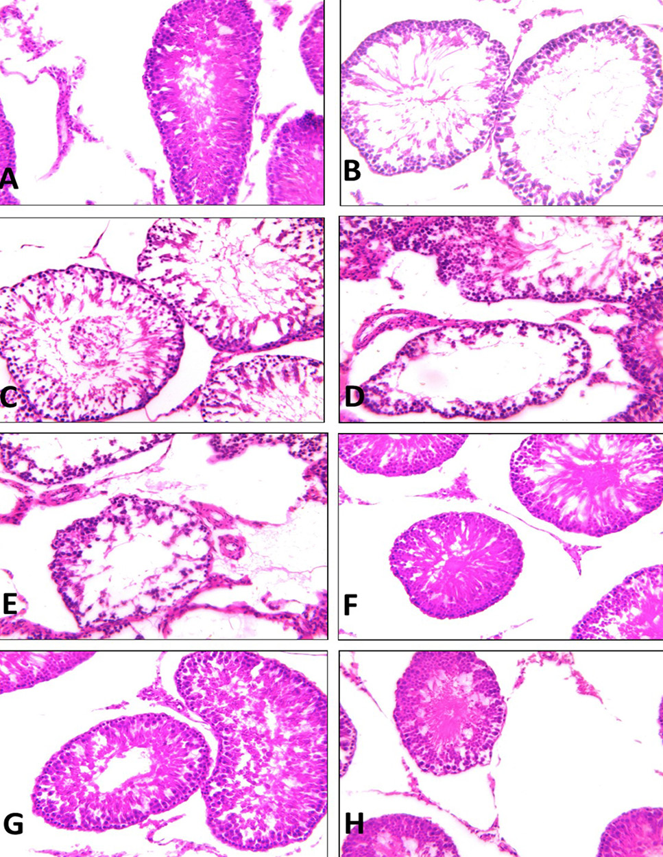
(A) Testis of control rats showing the normal histological structure of seminiferous tubules with different series of active spermatogenic layers, spermatozoa and the interstitial tissues. (B) Testis of mint treated group showing alternations of some tubules in the form of reduced the number of layers of the germinal epithelium, spermatozoa with vacuolated spermatogenic cells. (C, D and E) Testis of AL, AM and AH group showing tubular shrinkage with extensive degeneration of the germinal epithelium. The shrinkage tubules contained degenerated Sertoli cells with few germ cells. (F, G and H) Testis of ALM, AMM and AHM group showing no prominent histological changes. Most of the seminiferous tubules appeared to increase of spermatogenic cells and an increase in the number of sperm bundles was seen. (Stain HX and magnification x200).

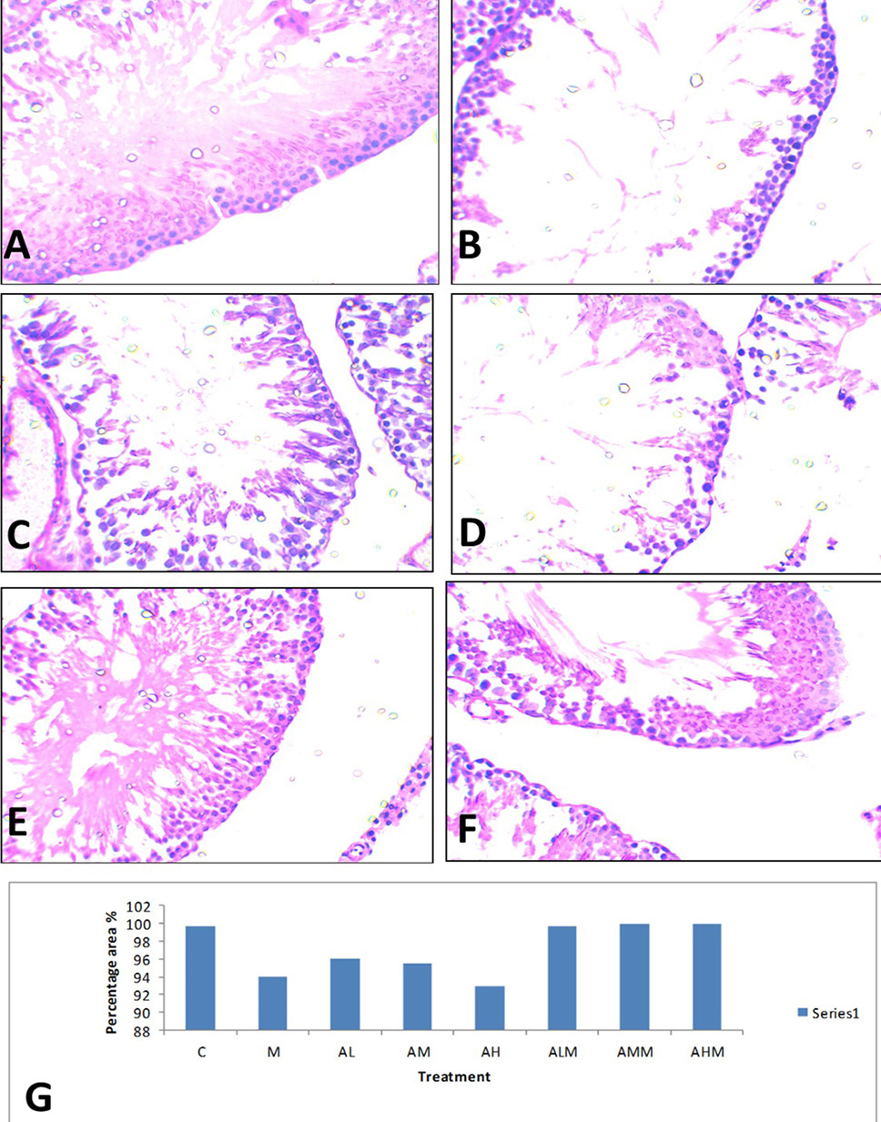
Cross sections from testis, (A) control group with positive PAS increase in the basement membrane of the tubules (the tunica albuginea) as well as in the intertubular connective tissue of the testes. (B, C, D and E) Testis of mint, AL, AM and AH treated group showing decrease PAS reaction. (F, G and H) Testis of ALM, AMM and AHM group showing normal positive PAS. (PAS staining technique, 400X). (I) Percentage of PAS stained area in the treated group.
Cross sections from testis, (A) control group with positive Feulgen reaction in the nucleus of spermatogonia and spermatocytes. (B, C, D and E) Testis of mint, AL, AM and AH treated group showing a decrease in The intensity of DNA blue color. (F, G and H) Testis of ALM, AMM and AHM group showing normal positive Feulgen reaction. (Feulgen staining technique, 200X).
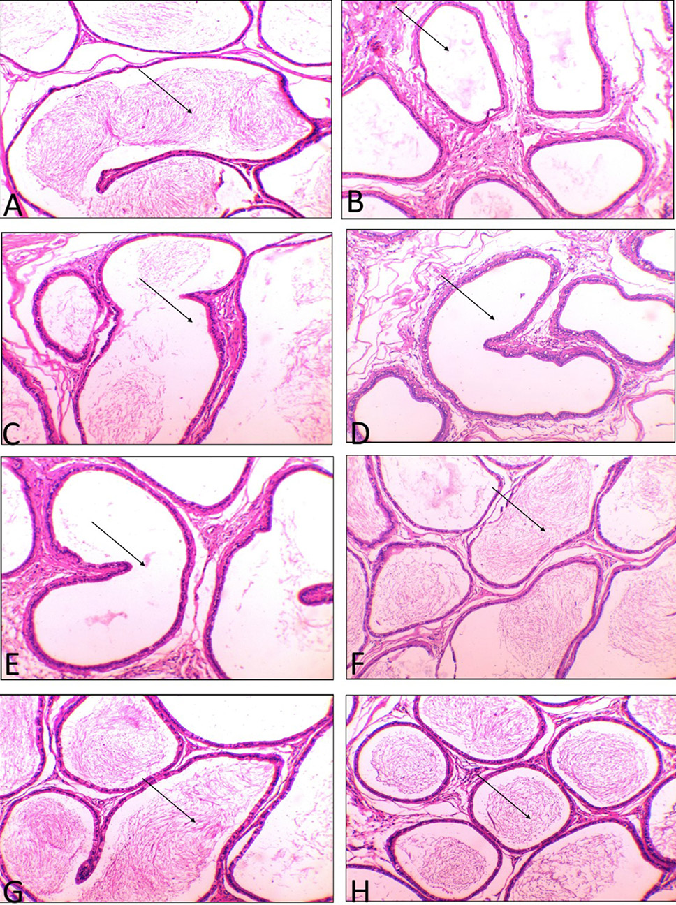
(A) Tubuli recti of the control group showing the normal histological structure lining with cuboidal epithelium and present of sperm in the lumen (←). (B) Tubuli recti of mint treated group showing alternations of some tubules in the form of reduced sperm in the lumen (←). (C, D and E) Tubuli recti of AL, AM and AH group showing vacuolation of the cuboidal epithelium with extensive absent of the sperm in the lumen (←). (F, G and H) Tubuli recti of ALM, AMM and AHM group showing no prominent histological changes with increase of sperm in the lumen (←). (H and E, x200).

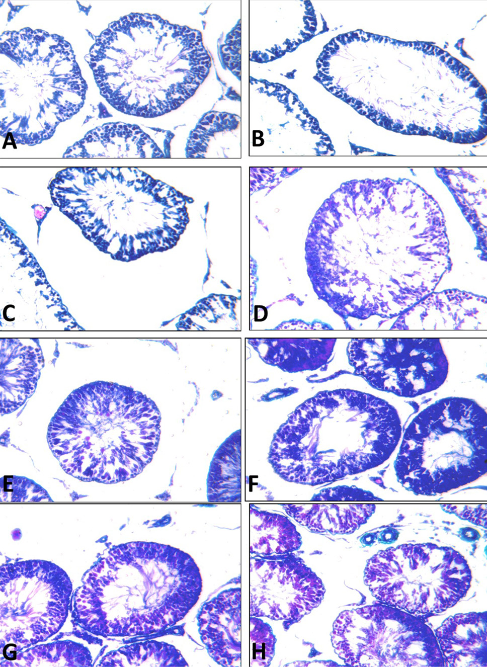
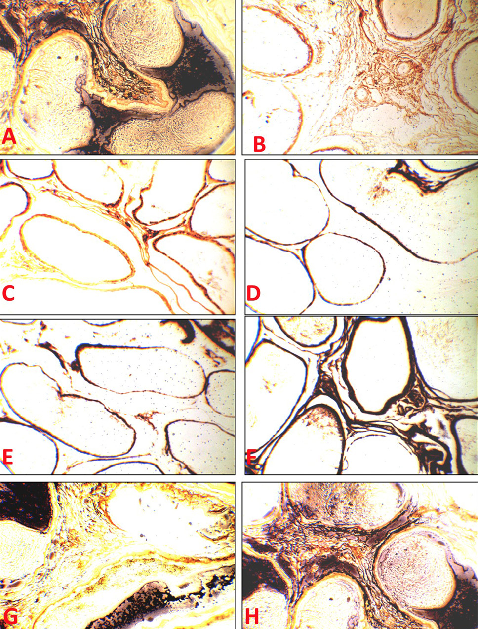
(A) Tubuli recti of the control group showing normal reticular fibers. (B, C, D and E) Tubuli recti of M, AL, AM and AH group showing degeneration and fragmentation of reticular fibers (F, G and H) Tubuli recti of ALM, AMM and AHM group showing the normal appearance of reticular fibers. (Silver stains x200).


