

Cellular Localization of MAPK, NF-κB and Nrf2 Signaling Pathways-Related Proteins in Crayfish Hepatopancreas


Cellular Localization of MAPK, NF-κB and Nrf2 Signaling Pathways-Related Proteins in Crayfish Hepatopancreas
Keqiang Wei1*, Yue Wei2 and Changxia Song1
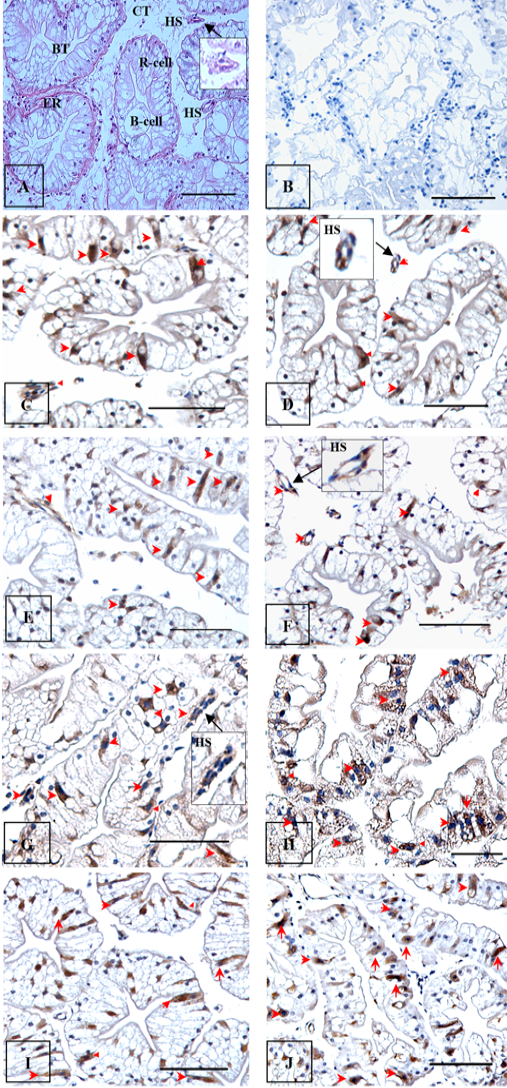
Localization and distribution of signaling pathway-related proteins in hepatopancreas of red swamp crayfish (P. clarkii). A, H and E staining; B, negative control (no primary antibody); C and D, p38MAPK; E and F, p-p38 MAPK; G and H, RelA (p65); I and J, Nrf2. The representative immunopositive cells were indicated with red arrowheads. Square indicated partial enlarged view. B-cell, secretory cell; BT, blind tubule; CT, connective tissue; ER, epithelium; HS, haemolymph sinusoids; R-cell, absorptive cell. 100 ×; scale bars=50 μm.

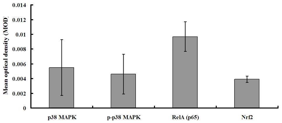
Tissue distribution of signaling pathway-related proteins in hepatopancreas of red swamp crayfish (P. clarkii). Expression levels were quantified as mean optical density (MOD) value of the immunopositive cells. Data were expressed as the mean ± SD (n = 6).


