

Atresia and Apoptosis in Pre- and Postovulatory Follicles of Sharptooth Catfish (Clarias gariepinus, Burchell, 1822)


Atresia and Apoptosis in Pre- and Postovulatory Follicles of Sharptooth Catfish (Clarias gariepinus, Burchell, 1822)
Şehriban Çek Yalnız 1,* and Erdal Yilmaz2

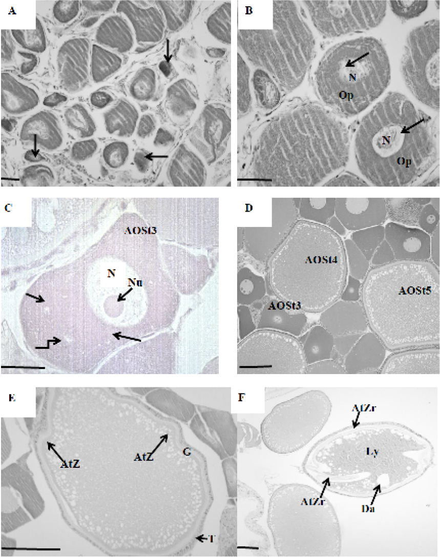
Pre-ovulatory atretic ovarian follicles. A, arrows show atretic oocytes as dark stain mass in the chromatin nucleolar stage; B, atretic oocytes at the perinocleolar stage, arrows show the development of clear spaces in the peripheral ooplasm; C, arrows show ealy vacuole formation; D, early phase of vascuolization, oocytes resorption; E, early degeneration of zona radiata in vitellogenic oocyte; F, fregmentation of zona radiata, follicular cells with protrusion extending into the oocyte cytoplasm. Ly, liquefaction of yolk granules; Da, degeneration of cortical alveoli; T, theca; G, granulosa; AtZr, atretic zona radiata. Scale bars: A, 30µm; B-F, 60 µm. Stain: hematoxylin & eosin.
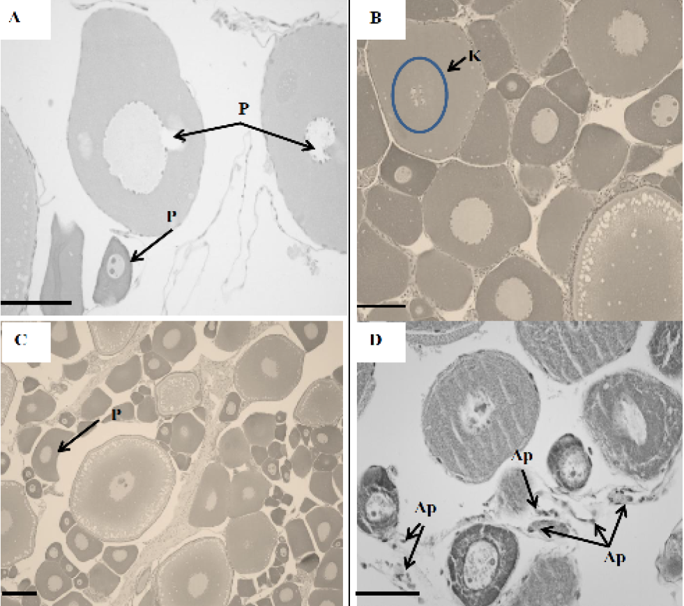
Apoptosis in pre-ovulatory follicles of C. gariepinus. A and C, arrow shows pyknosis, irreversible condensation of chromatin in the nucleus and condensation of cytoplasm of oocytes undergoing apoptosis; B, pyknosis followed by the fragmentation of the nucleus. (karyorhexis) and the budding phenomenon detected in pre-ovulatory oocytes of C. gariepinus. Arrow shows the budding phenomenon. P, pyknosis; K, karyorhexis; Ap, apoptotic body. Scale bar, 200µm. Stain: hematoxylin & eosin.
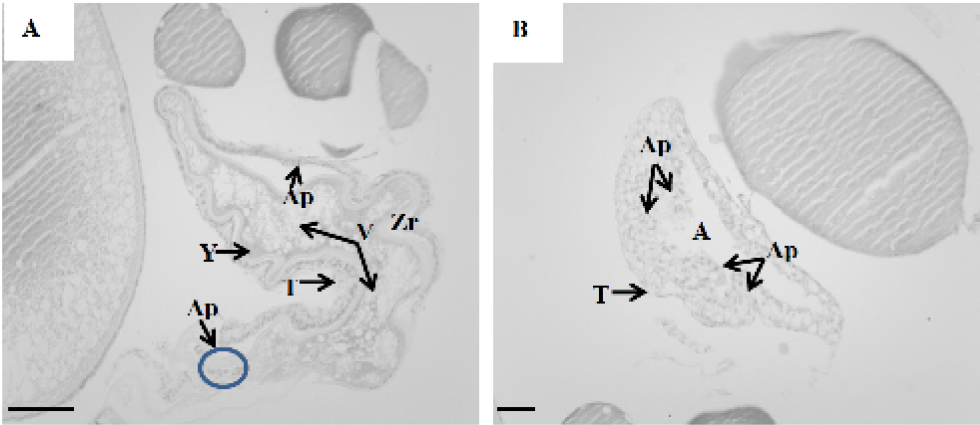
Apoptosis in pre-ovulatory follicles of C. gariepinus. A, engulfed yolk, disintegrated zona ratiata, hypertrophic theca cells, invading granulosa cells; B, apoptotic body formation is shown. Ap, apoptotic body; Y, yolk; V, vacuole; T, theca; Zr, zona radiata; A, atrium. Scale bar, 200µm. Stain: hematoxylin & eosin.

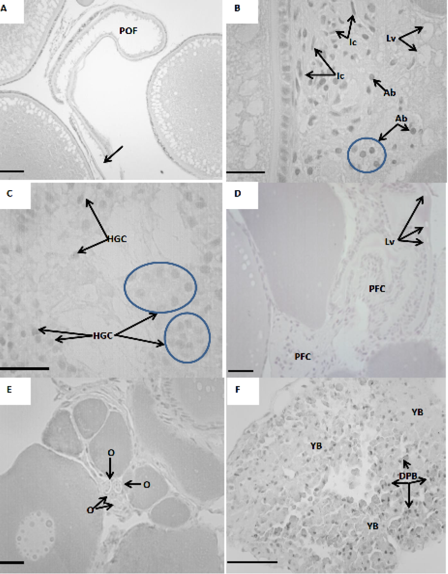
Atresia at post ovulatory follicle of C. Gariepinus. A, arrow shows evidence of the rupture of the follicular wall after ovulation; B, formation of interstitial cells; C, hypertrophy of granulosa cells are shown; D, phagocytıc follicular cells are shown; E, Arrow shows oogonia which were occasionaly detected amongst the connective tissue; F, arrows show yellow bodies which prominently represented in post ovulated ovaries. Dark pigmented bodies are also shown. POF, post ovulatory follicle; YB, Yellow bodies; Ab, apoptotic bodies; O, oogonia; DPB, dark pigmented bodies; HGC, hypertrophied granulosa cells; PFC, phagocytic follicular cells; Lv, large vacuole; Ic, interstitial cells. Scale bar, 200µm. Stain: hematoxylin & eosin.

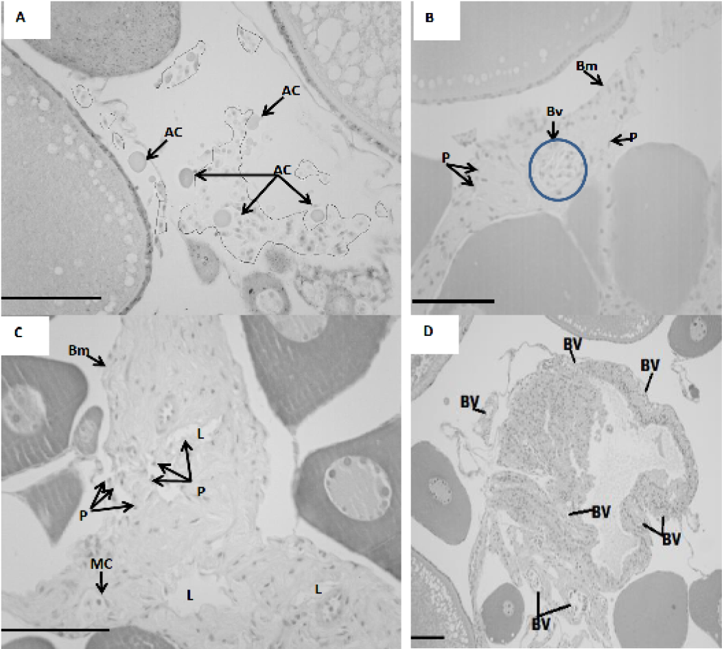
Apoptosis in POF. A, all irregular circulus show various type of blood cells. Arrows show apoptotic cells; B, arrow shows blood vessels. Piknosis were also detected; C, following inflection nuclei with condensed chromatin and showing pyknosis, were detected at the lumen of the follicle and at the follicular wall (arrows); D, blood vessels close to the follicular cells. Bm, basement membrane, MC, micropylar cell, AC, apoptotic cell; BV, blood vessels; P, pyknosis. Scale bars, 200µm. Stain: hematoxylin & eosin.


