

Quercetin Improves Liver Function, Decreases the Expression of Pro-apoptotic Proteins p53 and Bax and Increases the Antioxidant Defense of Hepatocytes in Aged Male Rats


Quercetin Improves Liver Function, Decreases the Expression of Pro-apoptotic Proteins p53 and Bax and Increases the Antioxidant Defense of Hepatocytes in Aged Male Rats
Eman A. Al-Shahari1,2, Eman R. ElBealy3, Abdelhalim A. Alkhazendar4 and Abeer A. Alm-Eldeen4*
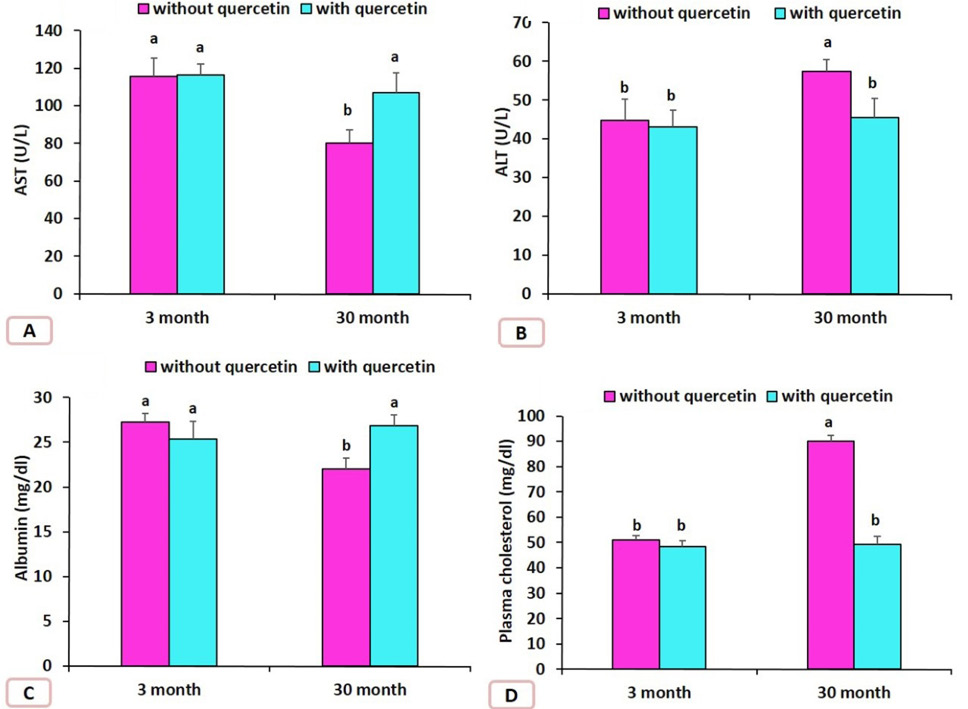
Effect of quercetin on the levels of AST, ALT, Albumin and plasma cholesterol in the rats that had 3 and 30 months old rats. Data are means ± SD with n=4, Different letters indicate significant differences among the columns (p ≤ 0.05).

Effect of quercetin on the liver MDA, SOD, CAT, GPx in the rats that had 3 and 30 months old. Data are means ± SD with n=4, Different letters indicate significant differences among the columns (p ≤ 0.05).

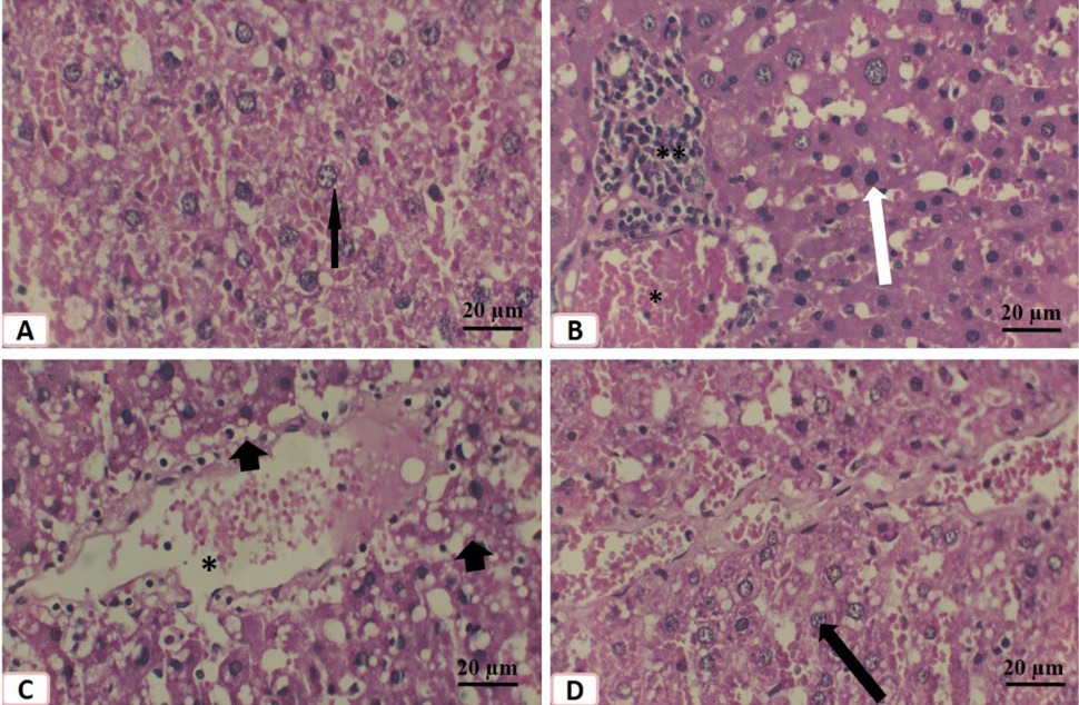
Effect of quercetin on liver of aged male rats. Histological structure of liver of 3 months old control rats (A), 3 months old treated liver (B), 30 months old saline treated rat (C) and 30 months old quercetin-treated rat (D). Note the vesicular nuclei (black arrows), cellular infiltration (**), dark nuclei (white arrow), vacuolated hepatocytes (black arrowhead) and congestion of the central vein (*). Stain: hematoxyline and eosin.
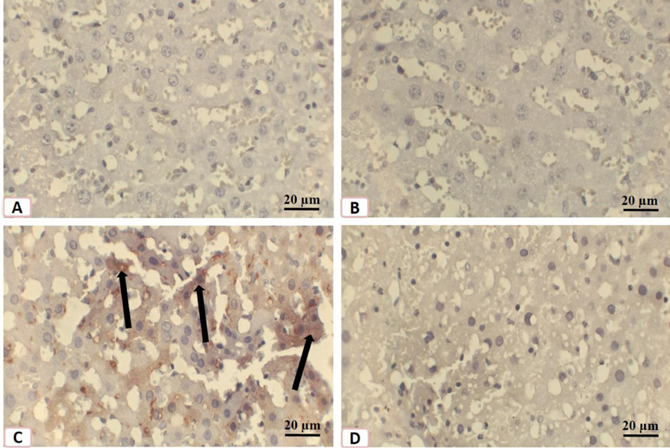
Immunostaining of histological sections of livers of rats with p53 antibodies. A, 3 months old control rats B, 3 months old quercetin-treated rats; C, 30 months old saline-treated rats; D, 30 months old quercetin-treated rats. Note the rarity of p53 expressions in A and B then the increase in its expression in C (arrows) then the rare of its expression in D.

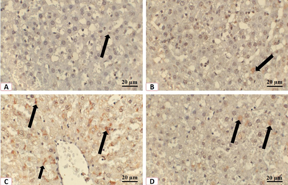
IImmunostaining of histological sections of livers of rats with bax antibodies. A, 3 months old control rat; B, 3 months old quercetin-treated rats; C, 30 months old saline-treated rats; D, 30 months old quercetin-treated rats. Note the rarity of bax expressions (arrows) in A and B then the increase of its expression in C (arrows) then the decrease of its (arrows) expression in D.


