

In vitro Assessment and Characterization of the Growth and Life Cycle of Leishmania tropica


In vitro Assessment and Characterization of the Growth and Life Cycle of Leishmania tropica
Qaisar Jamal*, Akram Shah, Syed Basit Rasheed and Muhammad Adnan

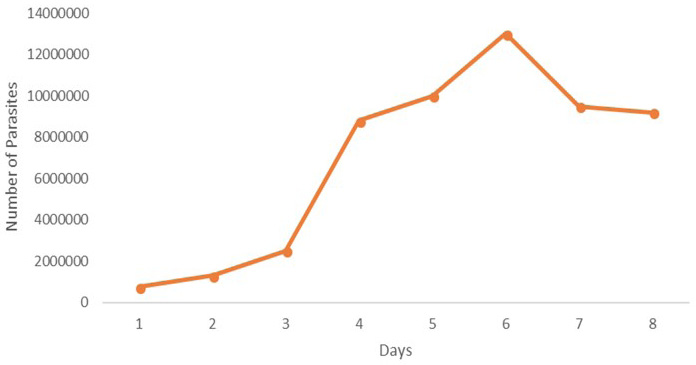
Growth curve of Leishmania tropica KWH23 over a week.

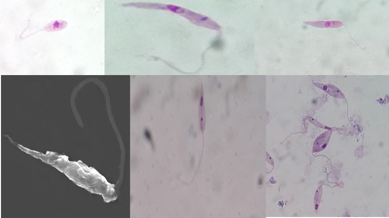
Different stages of L. Tropica KWH23 promastigotes in culture (See text for description).

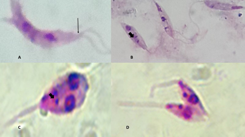
Division of the L. tropica promastigotes in axenic development. Cell with 2 flagella single kinetoplast and single nucleus (A), Cells with single flagellum, two kinetoplasts and single nucleus (B), Cell with Single flagellum, two kinetoplasts and two nuclei (C) and Cells with two flagella, two kinetoplasts and two nuclei near to the completion of cytokinesis (D). Solid arrows show kinetoplasts and the others indicate flagella.

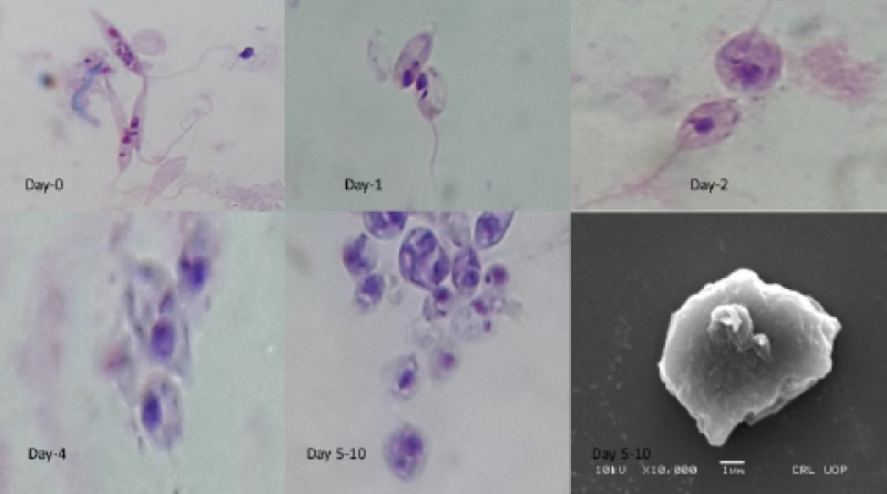
Promastigote to amastigote transformation. Change in the cell morphology and loss of flagellum is seen progressively from day 0-10. Lesion amastigotes are indicated by the arrow in the last inset.


