

Antifertility Efficacy of Testosterone on Male and Female Albino Mice


Antifertility Efficacy of Testosterone on Male and Female Albino Mice
Maha Mostafa1, Sohail Soliman1, Reham I. Mohamed2 and Wael M. El-Sayed1*

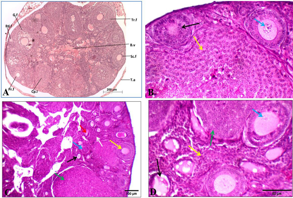
Effect of oral administration of exogenous testosterone on ovary of albino mouse. A shows histological structure of transverse section of the ovary of control female albino mice showing different stages of follicles. B.v: blood vessel, Cp.l: corpus luteum Pd. f: Primordial follicle, Pr. f: primary follicle, Sc. f: secondary follicle, G.f: Graffian follicle, T.a: tunica albuginea, Tr.f: tertiary follicle. B shows control ovary showing primary (black arrow) and secondary follicles with average oocyte (blue arrow), and corpus luteum (yellow arrow). C shows primordial (black arrow), primary (blue arrow), secondary follicles (yellow arrow), and multiple corpora lutea (green arrow) in cellular stroma (white arrow). D shows ovary from treated mouse showing primordial (black arrow) and secondary follicles with degenerated oocyte (blue arrow), and multiple corpora lutea (green arrows) in cellular stroma (yellow arrow). Stain: H and E. Magnification: A: 100X; B: 400X; C: 200X; D: 400X

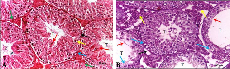
Effect of oral administration of exogenous testosterone on testes of albino rat. Histological structure of transverse section of testis of control male albino mouse showing A: average-sized seminiferous tubules (T) with average basement membranes (black arrow), spermatogonia (blue arrow), primary spermatocyte (yellow arrow), many sperms (red arrow) and average interstitium showing Leydig cells (green arrow). B: small-sized and dilated tubules (T) with thickened basement membrane (black arrow), thin germinal lining with scattered spermatogonia and primary spermatocytes with vacuolated cytoplasm (blue arrow), no sperms (red arrow), and excess interstitium with average Leydig cells (yellow arrow). Stain and Magnification H and E, X 400.


