

Antibiofilm Activity of Proteolytic Enzymes against Salmonella Gallinarum Isolates from Commercial Broiler Chickens


Antibiofilm Activity of Proteolytic Enzymes against Salmonella Gallinarum Isolates from Commercial Broiler Chickens
Iram Liaqat1*, Tahir Hussain1,2, Aisha Waheed Qurashi3, Gulbeena Saleem2, Asia Bibi4, Muhammad Fiaz Qamar5,ShaukatAli1 and Ikram-ul-Haq6

Biofilm quantifications of S. Gallinarum strains (SG 119/Pak, SG 421/ India and SG 2a 189/China) using test tube and liquid interface coverslip assays. (a). Three strains were grown in test tubes over a time period of 3, 5 and 7 days. Each set was taken out after respectives time period and stained with 0.1% CV stain. OD580 [/] was measured. (b). Three strains were grown on coverslips submerged in petriplates having nutrient broth. After day 1 and day 2, each of the respective set of coverslips was processed for biofilm quantification as mentioned in test tubes assay using CV staining method. Experiment was run in triplicates. One way ANOVA followed by post hoc Tukey test was used to analyze results in SPSS (Version 13.0). Bas with no common superscript are significantly different (P<0.05).

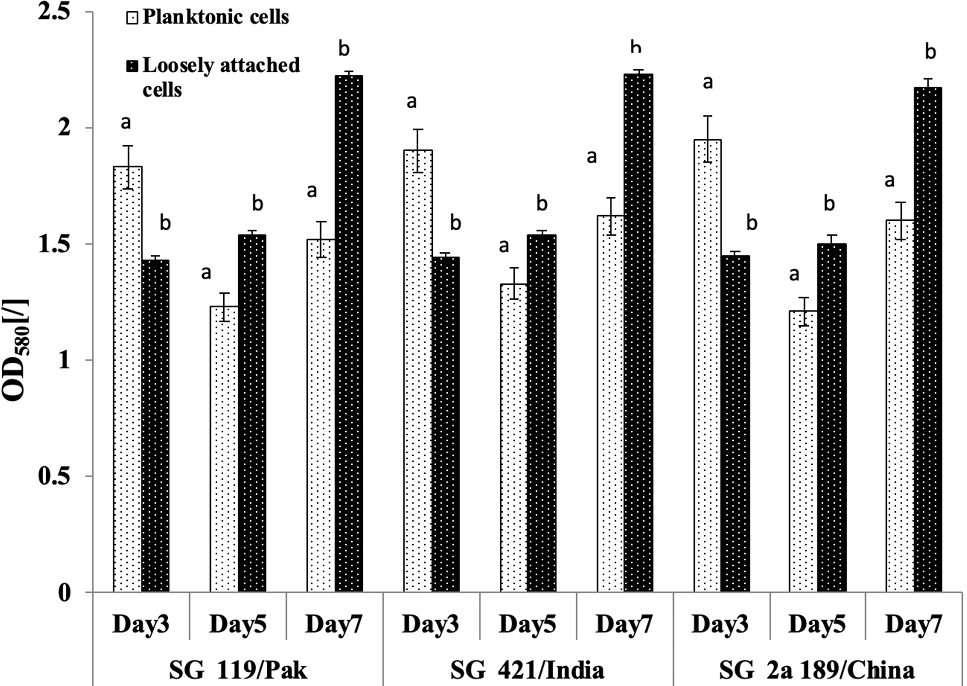
Quantitative determination of planktonic and loosely attached cells of three S. Gallinarum strains (SG 119/ Pak, SG421/India and SG 2a 189/China) using test tube assay via CV staining method. Briefly bacteria were grown in test tubes for 3, 5 and 7 days. Each set was taken out after respectives time period, cells were centrifuges and stained with 0.1% CV stain OD580 [/] was measured. Results are replica of three independent experiment. One way ANOVA followed by post hoc Tukey test was used to analyze results in SPSS (Version 13.0). Bars with no common superscript are significantly different (p<0.05).

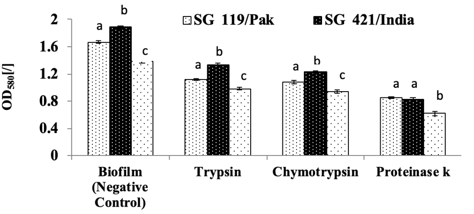
Effect of proteolytic enzymes (trypsin, chymotrypsin and proteinase k) on biofilm formation of three S. Gallinarum strains (SG 119/ Pak, SG421/India and SG 2a 189/China) using test tube assay via CV staining method. Briefly bacteria were grown in test tubes. 30mM PBS containing 5 X MIC of trypsin, chymotrypsin and proteinase k was added in each set of test tubes respectively. CV staining method was used to determine inhibitory the effect of enzymes on biofilm. Results are replica of three independent experiment. One way ANOVA followed by post hoc Tukey test was used to analyze results in SPSS (Version 13.0). Bars with no common superscript are significantly different (p<0.05).
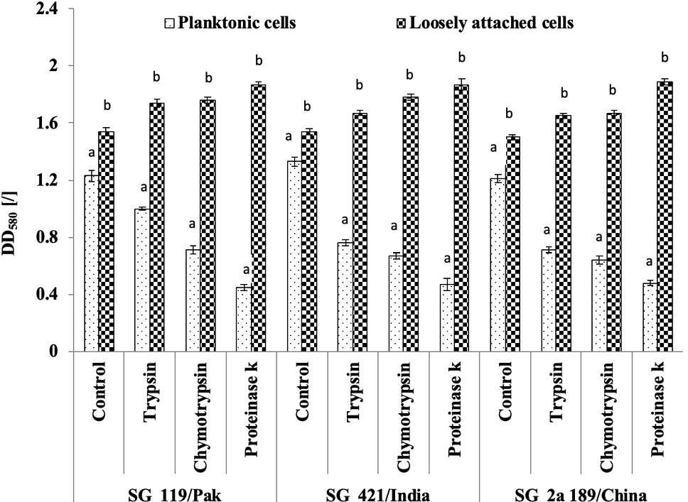
Effect of proteolytic enzymes (trypsin, chymotrypsin and proteinase k) on planktonic and loosely attached cells of three S. Gallinarum strains (SG 119/ Pak, SG421/India and SG 2a 189/China) using test tube assay via CV staining method. Briefly bacteria were grown in test tubes. 5 X MIC of trypsin, chymotrypsin and proteinase k were added in each set of test tubes respectively. CV staining method was used to determine inhibitory the effect of enzymes on planktonic and loosely attached cells. Experiment was run in triplicates. One way ANOVA followed by post hoc Tukey test was used to analyze results in SPSS (Version 13.0). Bars with no common superscript are significantly different (p<0.05).


